Late Jurassic pterosaur under UV light

Mike Eklund, Research Associate at the University of Texas,
is using black light (= uv light) to reveal what is ‘hidden in plain sight’ in fossils. At the HMNS.org blogsite a fully articulated and excellently preserved Late Jurassic pterosaur serves as only one of many subjects using this lighting technique (Figs. 1–3).

In case some controversial items are overlooked,
as they have been for centuries, I thought I’d highlight a few observations (Figs. 1-3).

Figure 1. Coloring the bones and membranes of this pterosaurs helps identify them here.

Figure 1. Coloring the bones and membranes of this pterosaurs helps identify them here.

For paleo-artists
note how the wing finger folds completely against the forearm. Note how the membrane virtually disappears when folded (Peters 2002, 2009), especially so at the wing tip. Also note that no part of the wing membrane ever extends to the tibia or ankle. This is evidence to counter myths perpetuated by prior pterosaur workers and artists.

Figure 2. Manual digit 5 on this pterosaur is undisturbed.

Figure 2. Manual digit 5 on this pterosaur is undisturbed.

Figure 3. Manual unguals on this pterosaur are undisturbed.

Figure 3. Manual unguals on this pterosaur are undisturbed.

This is not the first time
wing unguals and manual digit 5 have been identified in pterosaurs. Use those keywords to find previously posted specimens. Traditional paleontologists believe these bones don’t exist. That’s why I use Photoshop and the DGS technique… to share evidence. Now I encourage you to see for yourself.


References
Peters D 2002. A New Model for the Evolution of the Pterosaur Wing – with a twist. – Historical Biology 15: 277–301.
Peters D 2009. A reinterpretation of pteroid articulation in pterosaurs. Journal of Vertebrate Paleontology 29:1327-1330.

https://blog.hmns.org/2020/01/hidden-in-plain-sight-how-photography-techniques-are-helping-us-dig-deeper/


Preondactylus skeleton model on a tree

Rummaging through my file cabinets,
I ran across some Polaroid photos of a wire and putty model of Preondactylus (Figs. 1, 2) I made decades ago and mounted to a backyard branch. Note the sprawling femora, a lepidosaur trait.

Figure 1. Years ago, back in the days of Polaroid cameras, I built this to scale model of Preondactylus, mounted it on a tree branch and took its picture.

Figure 1. Years ago, back in the days of Polaroid cameras, I built this to scale model of Preondactylus, mounted it on a tree branch and took its picture.

Preondactylus bufarinii (Wild 1984, Dalla Vecchia 1998; Norian, Late Triassic, ~205 mya) was considered by Unwin (2003) to be the most basal pterosaur. It is not. Derived from a sister to the Italian specimen of AustriadactylusPreondactylus phylogenetically preceded Dimorphodon. Distinct from Austriadactylus, the skull of Preondactylus was lower and narrower with a larger antorbital fenestra completely posterior to the naris. The cervicals were shorter, the caudals more robust. The scapula and coracoid were more robust and straighter. The sternum was much larger. The humerus was anteriorly concave. The ulna and radius were shorter. The pelvis and pes were relatively longer. Pedal digit IV was shorter and V was longer. The metatarsals were longer than the pedal digits and IV was shorter than III.

Figure 2. At the time I thought I would use this photo of Preondactylus for a basis for an illustration with all the problems of perspective worked out.

Figure 2. At the time I thought I would use this photo of Preondactylus for a basis for an illustration with all the problems of perspective worked out. This is literally a ventral view.

Contra traditional pterosaur paleontologists,
who readily admit they have no idea which taxa are proximal outgroups to Pterosauria, basal pterosaurs, like Late Triassic Preondactylus and their fenestrasaur ancestors, were bipedal. Even so they continued to use their long, sharp-clawed free fingers to cling to trees like this (Figs. 1, 2).

Note the digitigrade pedes in this basal pterosaur,
distinct from the flat-footed beachcombers that made most of the tracks. By the way, we have tracks of digitigrade anurognathid pterosaurs (Peters 2011) derived from digitigrade dimorphodontids, like Preondactylus.

Earlier
here, here and here we looked at other ways pterosaurs could stand on and hold on to tree branches. Two of the many ways we know pterosaurs are lepidosaurs are the elongate manual digit 1 and the elongate pedal digit 5, neither of which appear in archosaurs, both of which appear in tritosaur lepidosaurs.

References
Peters D 2000a. Description and Interpretation of Interphalangeal Lines in Tetrapods. Ichnos, 7: 11-41.
Peters  D 2000b. A redescription of four prolacertiform genera and implications for pterosaur phylogenesis. Rivista Italiana di Paleontologia e Stratigrafia 106: 293-336
Peters D 2002. A New Model for the Evolution of the Pterosaur Wing – with a twist.
Historical Biology 15: 277-301.
Peters D 2007. The origin and radiation of the Pterosauria. Flugsaurier. The Wellnhofer Pterosaur Meeting, Munich 27.
Peters D 2011. A catalog of pterosaur pedes for trackmaker identification.
Ichnos 18(2):114-141. http://dx.doi.org/10.1080/10420940.2011.573605
Wild R 1984. A new pterosaur (Reptilia, Pterosauria) from the Upper Triassic (Norian) of Friuli, Italy, Gortiana — Atti Museo Friuliano di Storia Naturale 5:45-62.

wiki/Preondactylus

Big pterosaurs: big or little wing tips

Earlier and below (Fig. 2) we looked at large and giant pterosaur wings comparing them to the largest flying birds, including one of the largest extant flying birds, the stork, Ciconia, and the extinct sheerwater, Pelagornis, the largest bird that ever flew.

FIgure 2. A basal pteranodotid, the most complete Pteranodon, the largest Pteranodon skull matched to the largest Pteranodon post-crania compared to the stork Ciconia and the most complete and the largest Quetzalcoatlus

FIgure 1. A basal pteranodotid, the most complete Pteranodon, the largest Pteranodon skull matched to the largest Pteranodon post-crania compared to the stork Ciconia and the most complete and the largest Quetzalcoatlus. Note the much reduced distal phalanges in the complete and giant Quetzalcoatlus, distinct from the Pteranodon species.

Today
we’ll look at how the largest Pteranodon (Figs. 1, 4) compares to much larger pterosaurs, like Quetzalcoatlus northropi (Figs. 1, 2) that have vestigial wingtips similar to those of the  much smaller flightless pre-azhdarchid, SOS 2428 (Fig. 3).

Note the tiny three distal phalanges
on the wing of the largest Quetzalcoatlus, distinct from the more typical elongate and robust distal phalangeal proportions on volant pterosaurs of all sizes. Much smaller definitely flightless pterosaurs, like SOS 2428, shrink those distal phalanges, too. That’s the pattern when pterosaurs lose the ability to fly.

Figure 2. Q. northropi and Q. sp. compared to Ciconia, the stork, and Pelagornis, the extinct gannet, to scale. That long neck and large skull of Quetzalcoatlus would appear to make it top heavy relative to the volant stork, despite the longer wingspan. Pteranodon and other flying pterosaurs do not have such a large skull at the end of such a long neck (Fig. 1). The longer wings of pelagornis show what is typical for a giant volant tetrapod, and Q. sp. comes up short in comparison.

Figure 2. A previously published GIF animation. Q. northropi and Q. sp. compared to Ciconia, the stork, and Pelagornis, the extinct gannet, to scale. That long neck and large skull of Quetzalcoatlus would appear to make it top heavy relative to the volant stork, despite the longer wingspan. Pteranodon and other flying pterosaurs do not have such a large skull at the end of such a long neck (Fig. 1). The longer wings of pelagornis show what is typical for a giant volant tetrapod, and Q. sp. comes up short in comparison.Today we’ll compare the wingspan of the largest Quetzalcoatlus to the largest and more typical Pteranodon species (Fig. 2).

Unfortunately
pterosaur workers refuse to consider taxa known to be flightless, like SOS 2428 (Peters 2018). It’s easy to see why they would be flightless (Fig. 3). Scaled to similar snout/vent lengths with a fully volant pterosaur like n42 (BSPG 1911 I 31) the wing length and chord are both much smaller in the flightless form.

Lateral, ventral and dorsal views of SoS 2428

Figure 3. Lateral, ventral and dorsal views of the flightless SoS 2428 (Peters 2018) alongside No. 42, a volant sister taxon.

Comparing the largest ornithocheirid,
SMNK PAL 1136, to the largest Pteranodon (chimaera of largest skull with largest post-crania in Fig. 4) shows that large flyers have elongate distal phalanges, distinct from body and wing proportions documented in the largest azhdarchids, like Quetzalcoatlus.

Figure 5. Largest Pteranodon to scale with largest ornithocheirid, SMNS PAL 1136.

Figure 4. Largest Pteranodon to scale with largest ornithocheirid, SMNS PAL 1136. Note the long distant wing phalanges on both of these giant flyers. This is what pterosaurs evolve to if they want to continue flying. And this is how big they can get and still fly. Giant azhdarchids exceed all the parameters without having elongate wings. Note: the one on the left has a longer wingspan whir the one on the right has a more massive torso and skull together with more massive proximal wing bones and pectoral girdle. On both the free fingers are tiny, parallel oriented laterally and slightly tucked beneath the big knuckle of the wing finger. The pteroid points directly at the deltopectoral crest. 

As the largest Pteranodon and largest ornithocheirid (SMNS PAL 1136)
(Fig. 4) demonstrate, as flying pterosaurs get larger, they retain elongate distal wing phalanges. And big, robust phalanges they are.

By contrast in azhdarchids and pre-azhdarchids
there is a large size bump after n42 (BSPG 1911 I 31) the fourth wing phalanx either disappears (see Microtuban and Jidapterus) or shrinks to a vestige. Then there’s Zhejiangopterus (Fig. 5), with a big pelvis, gracile forelimbs and a giant skull on a very long neck. Just that neck alone creates such a long lever arm that the pterosaur is incapable of maintaining a center of balance over or near the shoulder joints.

Figure 1. Click to enlarge. There are several specimens of Zhejiangopterus. The two pictured in figure 2 are the two smallest above at left. Also shown is a hypothetical hatchling, 1/8 the size of the largest specimen.

Figure 5. There are several specimens of Zhejiangopterus. The two pictured in figure 2 are the two smallest above at left. Also shown is a hypothetical hatchling, 1/8 the size of the largest specimen.

As mentioned earlier, becoming flightless permitted, nay, freed azhdarchid pterosaurs to attain great size. They no longer had to maintain proportions that were flightworthy. Instead they used their shortened strut-like forelimbs to maintain a stable platform in deeper waters. And when they had to move in a hurry, their wings could still provide a tremendous amount of flurry and thrust (Fig. 6) for a speedy getaway.

Quetzalcoatlus running like a lizard prior to takeoff.

Figure 6. Quetzalcoatlus running without taking off, using all four limbs for thrust. That long lever arm extending to the snout tip in front of the center of gravity is not balanced in back of what would be the center of lift over the wings

For the nitpickers out there…
some specimens of Nyctosaurus (UNSM 93000, Fig. 7) also have but three wing phalanges, but they are all robust. The distal one is likely the fourth one because it remains curved. Phalanges 2 and 3 appear to have merged, or one of those was lost. Compare that specimen to a more primitive Nyctosaurus FHSM VP 2148 with four robust wing phalanges.

Figure 5. Cast of the UNSM 93000 specimen of Nyctosaurus. Missing parts are modeled here.

Figure 5. Cast of the UNSM 93000 specimen of Nyctosaurus. Missing parts are modeled here.

References
Peters D 2018. First flightless pterosaur (not peer-reviewed). PDF online.


Flugsaurier 2018: Los Angeles County Museum

Flugsaurier
is a meeting of those interested in pterosaurs that happens in another part of the world every few years. I went to the first few. Saw a lot of specimens. Met a lot of colleagues. Produced a few abstracts and gave some presentations.

Over the next few days
there’s a Flugsaurier meeting taking place in Los Angeles. Many well-known and not-so-well known speakers are giving presentations this year. I will not be among them. Why?

So far as I know,
all of the conveners and many of the presenters continue to ignore a paper I wrote 18 years ago on the origin of pterosaurs from fenestrasaurs, not archosaurs. Other papers followed on wing shape, trackmaker identification and other topics, all supporting that phylogenetic hypothesis of relationships. Evidently workers would prefer to hope that pterosaurs arose from archosaurs close to dinosaurs. This is not where the data takes anyone interested in the topic who is not a party to taxon exclusion.

In addition, several of the conveners

  1. subscribe to the invalid quad-launch hypothesis
  2. the bat-wing reconstruction of the brachiopatagium.
  3. they believe that pedal digit 5 framed a uropatagium.
  4. They refuse to add tiny Solnhofen pterosaurs to their cladograms.
  5. They refuse to add several specimens of each purported genus to cladograms—and because of this they don’t recognize the four origins of the pterodactyloid-grade (not clade).
  6. They still don’t recognize that pterosaurs grew isometrically.
  7. They still don’t accept that pterosaur mothers retained their egg/embryo within the body until just before hatching (a lepidosaur trait).
  8. They still don’t accept that pterosaur bone fusion patterns follow lepidosaur, rather than archosaur patterns.
  9. They accept the idea that giant eyeballs filled the anterior skulls of anurognathids, not realizing that the supposed ‘scleral ring’ on edge of the flathead anurognathid is actually the mandible and tiny teeth.
  10. They reject any notion that all basal and some derived pterosaurs were bipedal, despite the footprint and morphological evidence proving bipedal locomotion.
  11. They all hold out hope that the largest azhdarchids could fly.
  12. I was going to say that all workers believe that crest size and hip shape identify gender, when the evidence indicates these are both phylogenetic markers, but then I found an abstract in 2018 that casts doubt on the gender/crest/pelvis hypothesis. So there’s hope.

That’s a fairly long list of ‘basics’
that most pterosaur workers ‘believe in’ despite the fact that there is no evidence for these false paradigms — but plenty of evidence for the lepidosaur origin of pterosaurs, from which most of the above hypotheses follow.

I am not attending Flugsaurier 2018
because the convening pterosaur workers deny and suppress the data listed above. Plus, I can more actively and thoroughly test assertions made during the conference from ‘my perch’ here in mid-America.

Good luck to those attending. 
Test all assertions and hypotheses, no matter their source.

Largest ‘flying reptile’ from the Crato formation? Maybe not.

Cheng et al. 2018
report on a partial wing finger (MPSC R 1221, Fig. 1) that they say represents, “The largest flying reptile from the Crato Formation, Lower Cretaceous, Brazil.”

But is it? 

Figure 1. The as yet undescribed SMNS PAL 1136 specimen is much larger than comparable bones in the new specimen, MPSC R 1221.

Figure 1. The as yet undescribed SMNS PAL 1136 specimen is much larger than comparable bones in the new specimen, MPSC R 1221. If the scale bars are correct, the SMNS specimen is much larger.

No…
if the scale bars are correct. The larger, as yet undescribed, and very impressive SMNS PAL 1136 specimen (Fig. 1) is not mentioned in the text. I do not know if the SMNS specimen is from the Crato or Roualdo formation (I have not gone back to look up that datum). In any case, the authors overlooked this specimen, because it is not mentioned in the text or charts that list a few dozen other large pterosaurs. It should have been included. Of course, then the headline would have read, “…second largest…” and no one wants that.

So was this oversight intentional?
We’ll never know. The SMNS specimen has been in the literature for 24 years (Frey and Martill 1994).

Addendum several days later
The Crato Formation was not erected until 13 years after the 1994 paper by Martill, Bechly and Loveridge. Therefore all layers were considered Santana Formation in 1994. So the SMNS specimen from the Santana formation might have come from the upper or lower layers. It should have been included in the 2018 survey.

The authors conclude
“Based on the fusion of the extensor tendon process and the first wing phalanx and bone histology, MPSC R 1221 presents a subadult individual of a late ontogeny stage (OS5) at time of death, whichmeans that the final maximized wingspan might have been larger. This is corroborated by the osteohistological sections since this individual did not present an external fundamental system.” Look how eager the authors are to hang on to that superlative, ‘largest’, even though we know of at least one that is so much larger.

The authors do not realize
or continue to deny data, that pterosaurs do not follow archosaur fusion patterns during ontogeny—because pterosaurs are not archosaurs, and their fusion patterns follow phylogenetic patterns.

I never heard the term,
“external fundamental system” before. So, I looked it up: “A closely spaced series of lines of arrested growth that is called the External Fundamental System (EFS) indicates that adult size has been reached.” Now we all know!

I hope this blog post
will one day turn out dozens of young paleontologists who will read every paper they see with a seasoned and skeptical eye. If so, a few of you may someday become editors of academic journals or manuscript referees. When that happens, don’t let mistakes like this slip out. Having a website, like ReptileEvolution.com, that is full of data and illustrations, makes it easy to fact-check superlative claims, like this one, with just a few clicks.

On that note:
here (Fig. 2) is a published illustration of a pterosaur wrist from Duque and Barret 2018 with labels that were a little mixed up with regard for the ulna and radius. The referees should have caught this.

Figure 1. From figure 9 from Duque and Barreto 2018 with corrections noted and digit 5 colorized

Figure 2. From figure from Duque and Barreto 2018 with corrections noted and digit 5 colorized. This mistake should have been caught by the authors and referees, not me.

References
Cheng X, Bantim RAM, Sayão JM, Kellner AWA, Wang X and Saraiva AAF 2018. The largest flying reptile from the Crato Formation, Lower Cretaceous, Brazil. Historical Biology. https://doi.org/10.1080/08912963.2018.1491567
Duque RRC and Barret AMF 2018. New exceptionally well-preserved Pterosauria from the lower Cretaceous Araripe Basin, Northeast Brazil. Cretaceous Research 10.1016/j.cretres.2018.05.004
Frey E and Martill DM 1994. A new Pterosaur from the Crato Formation (Lower Cretaceous, Aptian) of Brazil. Neues Jahrbuch für Geologie und Paläontologie, Abhandlungen 194: 379–412.

Anhanguera animation at the NHM (London)

This one started off with so much promise
as the animators at the National History Museum (NHM) in London assembled their version of the ornithocheirid pterosaur, Anhanguera, bipedally (Fig. 1), as you’ll see when you click on the video under ‘References’.

Figure 1. Animated by the NHM, Anhanguera is bipedal and flapping its literally oversize wings.

Figure 1. Animated by the NHM, Anhanguera is bipedal and flapping its literally oversize wings standing on oversize feet with an undersized skull and hyperextended elbows and unbalanced stance.

Unfortunately there were some morphology issues (compared in Fig. 2):

  1. wings too long
  2. sternal complex missing
  3. gastralia missing (but rarely preserved in ornithocheirids)
  4. feet way too big
  5. skull too small
  6. tail too short
  7. not sprawling
  8. free fingers too big
  9. wing fingers should tucked tight against elbows (in the same plane)
  10. one extra cervical
  11. anterbrachia too short and gracile
  12. elbows overextended (in Fig. 1)
  13. too much weight put on forelimbs, center of balance (wing root) should be over the toes
  14. Prepubes are extremely rare in ornithocheirds, but when present they are tiny, putter-shaped and oriented ventrally in line with the bent femora, not anteriorly
Figure 2. NHM Anhanguera compared to skeletal image from ReptileEvolution.com.

Figure 2. NHM Anhanguera compared to skeletal image from ReptileEvolution.com. There are at least 10 inaccuracies here. See text for list.

Also unfortunately, the video quickly devolved
to the invalid and dangerous quad launch, when (doggone it!) it was all set up to do a more correct and  much safer bird-like launch. The laws of physics and biomechanics are ignored here, but at least David Attenborough narrates.

Figure 3. NHM Anhanguera quad launch select frames.

Figure 3. NHM Anhanguera quad launch select frames. The laws of physics and the limitations of biomechanics are ignored here.

Attempts to convince readers and workers
that the quad-launch hypothesis cheats morphology and physics (as recounted here and at links therein) have so far failed. But I’m not giving up. So, if anyone has a connection to the NHM in London, please make this post available to alert them of their accidental foray into wishful thinking and inaccurate morphology.

References
National History Museum (NHM) in London

Axial rotation: fingers in pterosaurs, toes in birds

A somewhat recent paper by Botelho et al. 2015
looked at the embryological changes that axially rotate metatarsal 1 to produce a backward-pointing, opposable, perching pedal digit 1 (= hallux).

Hallux rotation phylogenetically
Botelho reports: Mesozoic birds closer than Archaeopteryx to modern birds include early short-tailed forms such as the Confuciusornithidae and the toothed Enantiornithes. They present a Mt1 in which the proximal portion is visibly non-twisted, while the distal end is offset (“bent”) producing a unique “j-shaped” morphology. This morphology is arguably an evolutionary intermediate between the straight Mt1 of dinosaurs and the twisted Mt1 of modern birds, and conceivably allowed greater retroversion of Mt1 than Archaeopteryx.”

“D1 in the avian embryo is initially not retroverted9, and therefore becomes opposable during ontogeny, but no embryological descriptions address the shape of Mt1, and no information is available on the mechanisms of retroversion.”

Figure 1. Pes of the most primitive Archaeopteryx, the Thermopolis specimen.

Figure 1. Pes of the most primitive Solnhofen bird, the Thermopolis specimen. This digit 1 never left the substrate.

In Tyrannosaurus,
(Fig. 2) the entire metatarsal 1 with pedal digit 1 is rotated just aft of medial by convergence. It’s not axially rotated. It’s just attached to the palmar side of the pes. This pedal digit 1 was elevated above the substrate.

Figure 2. The semi-retroverted pedal digit 1 of Tyrannosaurus rex in two views.

Figure 2. The semi-retroverted pedal digit 1 of Tyrannosaurus rex in two views. This digit 1 was elevated above the substrate.

In some birds
like the woodpecker, Melanerpes, and the unrelated roadrunner, Geococcyx, pedal digit 4 is also retroverted. Sorry, I digress.

Further digression
The axial rotation of pedal digit 1 in birds is convergent with the axial rotation of metacarpal 4 in Longisquama (Fig. 3) and pterosaurs. In both taxa the manus was elevated off the substrate and permitted to develop in new ways. Manual digit 4 never leaves an impression in pterosaur manus tracks… because it is folded, like a bird wing, against metacarpal 4. In Longisquama such extreme flexion is not yet possible.

Figure 1. Longisquama left and right manus traced using DGS then reconstructed (below). This is a very large hand for a fenestrasaur and manual digit 4 is oversized, as in pterosaurs.

Figure 3. Longisquama left and right manus traced using DGS then reconstructed (below). This is a very large hand for a fenestrasaur and manual digit 4 is oversized and the metacarpal is axially rotated, as in pterosaurs. Manual digit 5 is useless, but not yet a vestige. A pteroid is present, as in Cosesaurus. The coracoid is elongate as in birds. The sternum, interclavicle and clavicle are assembled into a single bone, the sternal complex, as in pterosaurs.

Note the lack of space between
the radius and ulna in Longisquama. This is what also happens in pterosaurs. It prevents the wrist from pronating or supinating, as in birds and bats… which means, the forelimb is flapping, not pressing against the substrate, nor grasping prey. That means all those images of Longsiquama on all fours are bogus. Now you know.

So now we come full circle
While the toes of birds axially rotate and the wing metacarpal of pterosaurs axially rotates, the forearms of birds, pterosaurs and Longisquama do not axially rotate. No one wants their wing to twist.

References
Botelho JF, Smith-Paredes D, Soto-Acuña S, Mpodozis J, Palma V and Vargas AO 2015. Skeletal plasticity in response to embryonic muscular activity underlies the development and evolution of the perching digit of birds. Article in http://www.Nature.com/Scientific Reports · May 2015 DOI: 10.1038/srep09840

Why do pterosaur workers ignore the most basic data?

I don’t know why,
but today’s leading pterosaur experts are actively ignoring the data from the last twenty years while inventing their own fanciful versions of what pterosaurs looked like (Fig. 1) – while claiming to be the latest word on the subject. Today we’ll be looking at a short paper from the latest Flugsaurier book by Hone, Witton and Martill 2017. And we’ll criticize the artwork that crystalizes their latest intentions. This is part 1.

For some reason
Hone, Witton and Martill like to show ancient cartoons that have little to no bearing on the present knowledge base (Fig. 1). I think it’s an English thing since most, if not all of the old engravings are indeed English in origin and easily lampooned. ‘See how far we’ve come!’, they seem to be saying. Doing so only takes up space that could otherwise go to competing current versions – which they want to avoid.

We’ve seen this
earlier when English professor D. Naish preferred to criticize work that preceded (= was not included in) ReptileEvolution.com. He employed cartoons made by others, rather than artwork that was actually posted on the website to show how bad the whole website was.

Evidently
It’s what they like to do. Someday, perhaps, they’ll look in a mirror and see some of the faults I present here… using their own artwork – which will soon enough joint their ancient engravings in a drawer full of foolish ideas they can draw upon in future decades.

Figure 1. Images from Hone, Witton and Martill 2017 showing the 'evolution' of our concept of Dimorphodon. Compare the latest color version to tracings of the several skeletons in figure 2.

Figure 1. Images from Hone, Witton and Martill 2017 showing the ‘evolution’ of our concept of Dimorphodon. Artists are credited in the text. Compare the latest color version to tracings of the several skeletons in figure 2. The long tail is based on a disassociated fossil probably belonging to a campylognathoid.

In figure 1
images of Dimorphodon through time are presented from Hone, Witton and Martill 2017.

  1. Rev. GE Howman 1829. Probably based on the headless holotype BMNH R1034 (Fig. 2). The authors labeled this as ‘monstrous’ when ‘inaccurate’, ‘fanciful’ or ‘medieval’ would do.
  2. Owen 1870. Probably based on the short-skull specimen, BMNH 41212 (Fig. 2), along with the disassociated tail specimen. The authors labeled this rendition as ‘ungainly, bat-like’. Odd word choice when among all the presented illustrations it is the one most like Witton’s 2017 version (#5).
  3. H Seeley 1901. Probably based on the long-skull specimen, BMNH PV R 1035 (Fig. 2) In the their comment Hone, Witton and Martill report, ‘progressive interpretation of D. macronyx as an erect-limbed quadruped’, but note that a biped interpretation was also offered. They thought it best not to show that possibility. 
  4. K Padian 1983. Probably based on the short-skull specimen, BMNH 41212 (Fig. 2). The authors report, ‘a highly active, bird-like digitigrade biped, a controversial interpretation that nevertheless symbolises the reinvention of pterosaurs in the late twentieth century.’ While there are minor issues associated with this figure (the orientation of fingers 1–3 and pedal digit 5, the over-extension of the metatarsophalangeal joint, the great length of the tail), it is the one that is most closely based on the skeleton (Fig. 2). BTW, when authors use the word, ‘controversial’ it usually means it does not fit their world view, but they have no evidence against it, nor any evidence to support their traditional hypothesis. 
  5. M Witton 2017. Not sure which skeleton this one is based on as it appears to have been done entirely freehand from memory and imagination. The authors report, ‘Modern interpretation of D. macronyx adult and speculative juveniles reflecting contemporary interpretations of pterosaur soft tissues, muscle development and ecology.’ Ahem…we’ll run through this illustration step-by-step below.
Figure 2. Images of Dimorphodon from ReptileEvolution.com. The tail attributed to Dimorphodon is shown in figure 3.

Figure 2. Images of Dimorphodon from ReptileEvolution.com. The tail attributed to Dimorphodon is shown in figure 3.

You know, you really can’t go wrong
when you strictly adhere to the bones (Figs. 2,3), soft tissue (Peters 2002) and footprints of the most closely related taxa (Peters 2011), which were made by digitigrade and bipedal pterosaur trackmakers. Unfortunately no such citations appear in this chapter. Those are purposefully omitted.

Dimorphodon model by David Peters

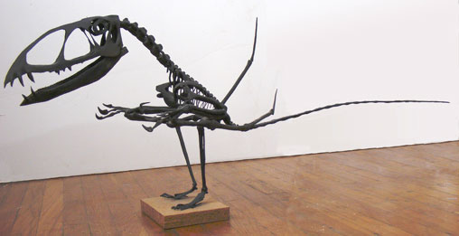
Figur 3. Dimorphodon model by yours truly. The tail is too long based on the disassociated tail.

Witton
fell under the spell of the quad-launch hypothesis (Habib 2008), then took it one step further and made Dimorphodon a galloping hunter (Fig. 4), forsaking its wings and erect, digitigrade hind limbs (according to related ichnite makers) to hunt prey on mossy logs with backward pointing fingers. The finger unguals are again too small here.

While writing this I became aware
of Sangster 2003, a PhD thesis that evidently used computer modeling to show Dimorphodon was a quadruped. I have not seen the thesis and Ms. Sangster can no longer be found online. I wonder about these conclusions because:

  1. PhD theses are, by definition, the work of inexperience workers; and
  2. Sangster may have had to earn her PhD by succumbing to the unveiled interests of her English professors, as we’ve seen before here and here.
Figure 4. Galloping Dimorphodon by Mark Witton.

Figure 4. Galloping Dimorphodon by Mark Witton.

To counter the awkward, dangerous and ultimately unproductive
quad-launch scenario, I proposed the following bipedal launch animation (Fig. 5). It combines the hind limb leap with the first flap of the large wings to provide the maximum thrust at takeoff. In the Habib proposal, you don’t get that wing flap until later in the cycle – maybe too late in the cycle. The quad launch also depends on directing the force of liftoff through the fragile free fingers. They were not strong enough for that, especialy not when there is a better option available using giant muscles in the chest and pelvis. That’s why the sacrum is so strong, to act as a fulcrum on that long, heavy lever!

FIgure 5. Dimorphodon take off (with the new small tail).

FIgure 5. Dimorphodon take off (with the new small tail).

So let’s get back
to Witton’s cover illustration (Fig. 6), which they tout as our contemporary view of Dimorphodon. I will note several inaccuracies (below). See figures 2 and 3 for accurate tracings.

Figure 6. Touted as the contemporary view of Dimorphodon, this Mark Witton illustration suffers from several fancies and inaccuracies.

Figure 6. Touted as the contemporary view of Dimorphodon, this Mark Witton illustration suffers from several fancies and inaccuracies.

  1. No Dimorphodon as this shape of skull.
  2. Needs a longer neck.
  3. Free fingers should be long and the unguals much larger.
  4. Wing appears to be too short with a too narrow wing tip chord.
  5. Witton wants to connect the trailing edge membrane from wing tip to ankle (or lateral toe), but look at the tremendous stretch in the membrane when that happens. Seems to be getting dangerously close to the narrow-at-the-elbow wing design of Zittel, Schaller and Peters, which they want to avoid.
  6. Ouch! This is a set of elongate toe bones with butt metatarsophalangeal joints – where Witton breaks them. This is not a calcar (a novel ossification on bat ankles which enters the uropatagium). One one side of these lateral toes the wing membrane attaches. On the other side the uroropatagium attaches. This is not shown in any fossil! Related taxa, from Langobardisaurus to Sharovipteryx, to Tanystropheus, with this same sort of elongate toe morphology, do not dislocate their bones this way. See Peters 2000 for a description that fits Rotodactylus tracks.
  7. No pterosaur has a uropatagium. This comes from a misinterpretation of Sordes. Pterosaur do have paired uropatagia.
  8. The tail is too large. On the BMNH 41212 fossil the traditionally overlooked tail is very small (Figs. 2, 7) This is in accord with related anurognathids. An unassociated tail has been attributed to Dimorphodon (Fig. 5) but it is robust and much longer. It probably belongs to a eudimorphodontid or campylognathoid. I”m surprised the tiny tail of Dimorphodon has gone unnoticed for so long. The specimen has been in English storage for over a hundred years. It was their responsibility for discovering this, but they chose instead to use their imaginations (Fig. 6).
  9. No tail vane is known for Dimorphodon. Tail vanes are found in pterosaurs like Campylognathoides and Rhamphorhynchus, both with a robust tail. Vestigial tails are unlikely to have had tail vanes.
FIgure 7. The tail of Dimorphodon (BMNH 41212 specimen). See figure 2 for reconstruction.

FIgure 7. The tail of Dimorphodon (BMNH 41212 specimen). See figure 2 for reconstruction.

I’m asking my Engllsh colleagues
|to step up their game and become more professional. Otherwise chaps from across the pond are going to continue pointing out the flaws in their thinking. I’m not going to say their approach is not scientific (as they say about my work), but when you forsake accuracy for artistry, you’re treading very close to that line.

References
Habib MB 2008. Comparative evidence for quadrupedal launch in pterosaurs. Zitteliana B28:159-166.
Hone DWE, Witton MP and Martill DM 2017.
New perspectives on pterosaur paleobiology in Hone DWE, Witton MP and Martill DM (eds) New Perspectives on Pterosaur Palaeobiology. Geological Society, London, Special Publications, 455, https://doi.org/10.1144/SP455.18
Peters D 2000. Description and Interpretation of Interphalangeal Lines in Tetrapods 
Ichnos, 7: 11-41.
Peters D 2002. A New Model for the Evolution of the Pterosaur Wing – with a twist 
Historical Biology 15: 277-301
Peters D 2011. A Catalog of Pterosaur Pedes for Trackmaker Identification.
Ichnos 18(2):114-141. http://dx.doi.org/10.1080/10420940.2011.573605
Sangster S 2003. The anatomy, functional morphology and systematics of Dimorphodon macronyx (Diapsida: Pterosauria)..Unpublished PhD thesis, University of Cambridge.

First African pterosaur trackway (manus only)

FIgure 1. From Masrour et al. 2017, manus only pterosaur tracks. They are BIG!

FIgure 1. From Masrour et al. 2017, manus only pterosaur tracks. They are BIG! Again I will note, only lepidosaurs can bend their lateral metacarpophalangeal joints within the palmar plane at right angles to the others, producing posteriorly oriented manual digit 3.

Masour et al. 2017
bring us new manus only Late Cretaceous azhdarchid tracks. They report, “The site contains only manus tracks, which can be explained as a result of erosion of pes prints.” They assume that the pterosaur fingers pressed deeper, carrying more weight on the forelimbs. Of course, this is a bogus explanation. No tetrapods do this. Pterosaurs put LESS weight on their tiny fragile fingers. They used their hands like skiers used ski poles.

FIgure 2. From Masrour et al. 2017, model of the trackmaker of the manus only tracks.

FIgure 2. From Masrour et al. 2017, model of the trackmaker of the manus only tracks erroneously attributed to Bennett 1997, who drew Pterodactylus, not this generalized azhdarchid.

There is another explanation for manus only tracks
called floating and poling, but that hypothesis was dismissed by the authors.

Masrour et al. dismiss the possibility of floating
by referencing Hone and Henderston 2014 in which simulations of the buoyancy of poorly constructed pterosaurs made using computers indicate that these reptiles had no ability to float well in water. This hypothesis was dismantled earlier here. In addition, Hone’s track record is not good. Neither is Henderson’s, who does not seem to care about using accurate skeletal reconstructions.

More importantly,
if Hone and Henderson put forth an anti-floating hypothesis no one (and certainly no scientist) should simply believe in it. This is Science. Others, like Masrour et al., should TEST hypotheses for validity, as was done here. Instead Masrour et al. put forth a hypothesis in which pes tracks were selectively erased over time, which seems preposterous and unnatural. This sort of selective erasure has never been observed in Nature.

Figure 1. The azhdarchid pterosaur Quetzalcoatlus floating and poling producing manus only tracks.

Figure 3. The azhdarchid pterosaur Quetzalcoatlus floating and poling producing manus only tracks. Remember the skull is as light as a paper sculpture.

Scientists fail
when they blindly follow bad hypotheses, just because they are published. Nodding journalists repeat what they read, whether right or wrong. Scientists test whenever they can.

Figure 5. Tapejara poling while floating, producing manus-only tracks, all to scale.

Figure 4. Tapejara poling while floating, producing manus-only tracks, all to scale. Remember the skull is as light as a paper sculpture.

Don’t believe in Henderson cartoons
(Fig. 5). Test with accurate representatives of skeletons IFig. 4).

Computational models of two pterosaurs from Hone and Henderson 2013. Note how both have trouble keeping their nose out of the water. Henderson's models have shown their limitations in earlier papers.

Figure 5. Computational models of two pterosaurs from Hone and Henderson 2013/2014. Note how both have trouble keeping their nose out of the water. Henderson’s models have shown their limitations in earlier papers.

When you don’t use cartoons for data
then you have a much better chance of figuring out how Nature did things.

Figure 4. Two configurations for Rhamphorhynchus. Because the wings act like pontoons, the torso and skull can be rotated relative to the wings to adopt a variety of floating configurations. Also note the large webbed feet, preserved in the darkling specimen. The tail can be elevated at its base.

Figure 6. Two configurations for Rhamphorhynchus. Because the wings act like pontoons, the torso and skull can be rotated relative to the wings to adopt a variety of floating configurations. Also note the large webbed feet, preserved in the darkling specimen. The tail can be elevated at its base.


Thank you for your continuing interest.
After over 2000 blog posts the origin of bats continues to be the number one blog post visited week after week, with totals equalling the sum of the next five topics of interest. That’s where the curiosity of the public is right now.

References
Hone DWE, Henderson DM 2014. The posture of floating pterosaurs: Ecological implications for inhabiting marine and freshwater habitats. Palaeogeography, Palaeoclimatology, Palaeoecology 394:89–98.
Masrour M et al. (4 other authors) 2017. 
Anza palaeoichnological site. Late Cretaceous. Morocco. Part I. The first African pterosaur trackway (manus only). Journal of African Earth Sciences (in press) 1–10.


https://pterosaurheresies.wordpress.com/2013/12/06/pterosaurs-were-unlikely-floaters-hone-and-henderson-2013/

Pterodactylus manual digit 5

Tiny, vestigial manual digit 5
sits on the top of the giant axially rotated metacarpal 4 of all pterosaurs. Here (Fig. 1) manual digit 5 is curled up on this Pterodactylus scolopaciceps specimen (BSP 1937 I 18), a pregnant pterosaur. Photoshop helps this digit ‘pop’ making it harder to overlook. A reconstruction unrolls it.

Figure 1. Manual digit 5 on top of the giant metatarsal 4 on Pterodactylus. It's easy to overlook, until you look for it.

Figure 1. Manual digit 5 on top of the giant metatarsal 4 on Pterodactylus. It’s easy to overlook, until you look for it.

References
Broili F 1938. Beobachtungen an Pterodactylus. Sitz-Bayerischen Akademie der Wissenschaten, zu München, Mathematischen-naturalischenAbteilung: 139–154.
Wellnhofer P 1970. Die Pterodactyloidea (Pterosauria) der Oberjura-Plattenkalke Süddeutschlands. Abhandlungen der Bayerischen Akademie der Wissenschaften, N.F., Munich 141: 1-133.

wiki/Pterodactylus