Out of 3400 prior posts
only two prior posts focused on carpals. One looked at the prepollux (radial sesamoid) of pandas and the pteroid + preaxial carpal of pterosaurs. Two looked at whale carpals here.
At present
the large reptile tree (LRT, 1825+ taxa) includes relatively few carpal traits, and none related to the migration of the pisiform and carpal 4 in mammals (see below). Crocodylomorphs elongate the proximal carpals. Many taxa do not ossify the carpals. As mentioned above, fenestrasaur centralia migrate to become the pteroid and preaxial carpal in pterosaurs. So some carpals are more interesting than others.

Figure 1. (Left) Diplovertebron right manus dorsal view. Carpal elements colored. (Right) Thrinaxodon right manus dorsal view. Some elements rotated to fit reconstruction. Some phalanges are reduced to discs in Thrinaxodon on their way to disappearing in mammals.
I was also interested
in the origin of the styliform process on the human ulna. It is located where the pisiform is located in Diplovertebron (Fig. 1) a basal archosauromorph amphibian-like reptile. And thus began a look at sample taxa in the lineage of humans.
The next step
was the basal cynodont, Thrinaxodon (Fig. 1). Here the elements are larger, link closer to one another and are better ossified. Some phalanges are reduced to discs in Thrinaxodon on their way to disappearing in mammals.

Figure 2. Right manus of the platypus, Ornithorhynchus (left) and early therian, Eomaia (right). Carpal elements colored. Note the disappearance (or fusion) of distal tarsal 4 in Eomaia along with the centralia.
The next step in carpal evolution is represented by the basalmost mammal,
Ornithorhynchus (Fig. 2), the platypus. Here distal tarsal 5 is ventral to the lateral centralia. The pisiform is tiny. The radiale and ulnare completely cap the radius and ulna. The platypus is a highly derived monotreme, not a basal taxon.
The enlargement of the distal radius width
relative to the distal ulna width begins with Eomaia (Fig. 2), a basal therian. So does the enlargement of distal carpal 5, taking the place of distal carpal 4.
The migration of tiny distal 4 to the palmar surface
is documented in the evolution of human carpals (Fig. 4), but probably originated with Eomaia (Fig. 2) where distal tarsal 4 is not diagrammed.
At this point it is worth noting
that mammal carpals have different names than those of other tetrapods. Here are the mammal homologs (which we will ignore):
Proximal Tarsals:
-
- Radiale = Scaphoid (lavendar)
- Intermedium = Lunate (tan)
- Ulnare = Triquetrum (dull pink)
- Pisiform = Pisiform (yellow green)
Centralia
-
- Medial Centralia = Prepollex (blue gray)
- Lateral Centralia = Lateral Centralia (blue gray)
Distal Tarsals:
-
- DT1 = Trapezium (yellow)
- DT2 = Trapezoid (orange)
- DT3 = Capate, magnum (green)
- DT4+5 = Hamate, unciform (4= blue, 5=purple)

Figure 3. Right manus dorsal view of basal tree shrew, Ptilocercus (left), and basal lemur, Indri (right). Carpal elements colored.
The next step in carpal evolution is represented by a basal placental,
Ptilocercus (Fig. 3), a tree shrew close to the base of the gliding and flying mammals. The fusion of distal tarsal 3 to the medial centrale is seen in Ptilocercus and its descendants. The ulna has a styloid process and the pisitorm extends laterally. Distal tarsal 1 is medially elongate to support a diverging thumb, further supported by the medial centralia.
Turns out the styloid process of the ulna
is not a fused carpal, but a novel outgrowth of the distal ulna appearing in basal placentals. The styloid process may have something to do with the ability of basal placentals to laterally rotate the manus for tree climbing in any orientation, including inverted, and to create a stop to prevent further rotation. Bats take this ability to its acme during wing folding.

Figure 4. Manus of human (Homo) in dorsal (left) and ventral/palmar (right) views. Carpal elements colored. Carpal 4 and pisiform palmar only. Compare to Diplovertebron (Fig. 1) in which so little has changed, including relative finger length.
The final step in carpal evolution
takes us from the lemur, Indri (Fig. 3) to the human, Homo (Fig. 4). Here a ventral (palmar) view of the manus is also provided so we can finally see the ultimate destination of distal tarsal 4.
Before finishing this blog post
scroll back and forth between figures one and four to see how close the human hand and all of its proportions so greatly resembles that of a very basal ampibian-like reptile. Even the relative finger length is the same. This is probably the most important takeaway today. The LACK of change is the news story here. Dinosaurs, horses and snakes cannot make the same statement.
There is no reason to continue using
the mammal specific identification of the carpals in paleontology when those bones are homologs to tetrapod wrist bones going back to the Devonian. Medical communities should also start using tetrapod homologs and let the analog identities fade into history.
Simply put:
There are five distal carpals named one through five in tetrapods. Some of them fuse with other carpals. There are three centralia. Some of these fuse with other carpals. Tetrapods have three proximal carpals. Their names are easy. The radiale is on the radius. The ulnare is on the ulna. The intermedium is intermediate between them. These tend not to fuse with other carpals, at least in basal placentals. And finally the pisiform appears by itself on the lateral margin sometimes in contact with the distal ulna sometimes not.
On a similar note,
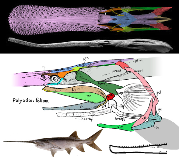
we supported earlier efforts to provide tetrapod homologs for fish skull bones here. Make things simple. There is enough hard work out there without needlessly translating bone identities.
References
Hamrick MW and Alexander JP 1996. The Hand Skeleton of Notharctus tenebrous (Primates, Notharctidae) and Its Significance for the Origin of the Primate Hand. American Museum Novitates 3182, 20pp.
Kielan-Jaworowska Z 1977. Evolution of the therian mammals in the Late Cretaceous of Asia. Part n. Postcranial skeleton in Kennalestes and Asioryctes. In: Z. Kielan-Jaworowska (ed.) Results Polish Mongolian Palaeont. Expeds. VIII. – Palaeont, Polonica, 37, 65-84.
Peters D 2009. A reinterpretation of pteroid articulation in pterosaurs. Journal of Vertebrate Paleontology 29:1327-1330.
Salesa MJ, Antón M, Peigné S and Morales J 2005. Evidence of a false thumb in a fossil carnivore clarifies the evolution of pandas. Proceedings of the National Academy of Sciences of the United States of America. abstract and pdf

