A new, tiny choristodere, Mengshanosaurus, enters the LRT

It’s tiny and probably a hatchling because sister taxa are much larger.
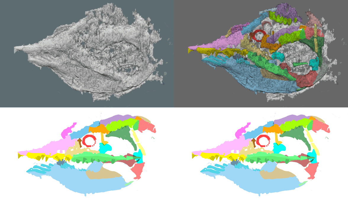
Mengshanosaurus minimus (Yuan et al. 2021; Early Cretaceous, China; skull length 3.5cm) nests between Ikechosaurus and Champsosaurus in the LRT (subset Fig. 3). Note the indented remnants of the antorbital fenestra in the hatchling model (Fig. 1). Apparently the post-frontal fontanelle is not a pineal opening. Sister taxa do not have a pineal opening.

Figure 1. Full scale model of CT-scanned Mengshanosaurus skull.

Traditional skull misinterpretations continue in Yuan et al.
Yuan et al did not properly label several fused bones (corrected Fig. 2 right) because they don’t know which taxa are choristodere outgroups and last common ancestors. That remains a traditional academic enigma that no one else seems to want to resolve, confirm or refute (Fig. 3) by simply adding taxa to find out.

Figure 2. Holotype of Mengshanosaurus.

Similarly,
the authors had no idea where to nest choristoderes as reptiles. In the large reptile tree (LRT, 1879+ taxa; subset from 2013 in Fig. 3) choristoderes nest as derived proterosuchids. Tiny transitional taxa, like the BPI 2871 specimen, lose the antorbital fenestra. Sister clades within the Pararchosauriformes include the Parasuchia and Proterochampsia. Euarchosauriformes derived from Euparkeria evolve to Archosauria, Rauisuchia, Erythrosuchia, etc.

Figure 3. Subset of the large reptile tree focusing on the pararchosauriformes and the Choristodera.
Figure 3. Subset of the large reptile tree from 2013 focusing on the pararchosauriformes and the Choristodera. This has not changed much, but for the addition of taxa, like Mengshanosaurus between Ikechosaurus and Champsosaurus.

If you don’t know where your clade resides,
keep adding taxa until it becomes apparent and all candidate sister taxa are considered. Or just sneak a peek at the LRT. Don’t overlook tiny taxa. Often tiny taxa bridge gaps, forming transitions at the genesis of major clades in a process known as phylogenetic miniaturization. This time a tiny taxon just turned out to be a hatchling.

Figure 4. The choristodere, Champsosaurus laramiensis (USNM PL 544147) has a vestige antorbital fenestra in the usual place, anterior to the orbit. Here the frontal fontanelle is also present, as in Mengshanosaurus.

PS
Sometimes adult choristoderes also retain a vestige of the antorbital fenestra (Fig. 4).


References
Yuan M, Li D-Q, Ksepka DT and Yi H-Y 2021.
A juvenile skull of the longirostrine choristodere (Diapsida: Choristodera), Mengshanosaurus minimus gen. et sp. nov., with comments on neochoristodere ontogeny. Vertebrata PalAsiatic in press DOI: 10.19615/ j.cnki.2096-9899.210607

wiki/Mengshanosaurus – not posted yet

“The amazing diversity of fishes” YouTube video

Dr. Phil Hastings,
Scripps professor and curator of the SIO Marine Vertebrate Collection delivers an online lecture and slide show sponsored by the University of California Television (UCTV). It runs for about an hour.

This is a traditional view of fishes
lacking any mention of fossil taxa.

Unfortunately
Hastings follows molecular data. Hastings mentions that we humans are indeed members of the Osteichthys. Ichthyologist Neil Shubin and artist Ray Troll are also mentioned.

By contrast,
the large reptile tree (LRT, 1836+ taxa) presents a distinctly different view of fish systematics because it includes fossils and minimizes taxon exclusion.

The razorfish, Aeoliscus, enters the LRT as an upside-down sea horse

Figure 1. Aeoliscus in vivo at full scale on a 72 dpi monitor.

Aeoliscus strigatus (Günther 1861, aka Centriscus strigatus, Amphisle strigata; 15cm) is the extant razorfish or shrimpfish. In the large reptile tree (LRT, 1834+ taxa) the shrimpfish nests with the sea horse, Hippocampus (Fig. 2). Thus Aeoliscus can be thought of as an upside-down sea horse despite many morphological difference (= no other tested vertebrate is closer). The dorsal fins have migrated to the caudal area. The caudal fin migrates to the anal area, bending the vertebral column to do so (Fig. 2). The tiny mouth, perhaps the smallest among vertebrates, is used like an eye-dropper to suck up minute brine shrimp. The preoperculum and operculum are large to produce suction for the tube mouth.

The preoperculum extends anteriorly beyond the orbit, merging with the quadrate (Fig. 2, at least in Gregory’s 1933 drawing). The lacrimal is absent (at least in Gregory’s 1933 drawing). As in the sea horse the naris is close to the orbit. The orbit is confluent with the antorbital fenestra. The supratemporal is absent or fused.

The shape of Aeoliscus was for hiding among sea grasses and other vertical sea floor environmental elements. The counter-shading of the body increases the illusion of slender background elements.

Figure 2. Aeoliscus anatomy from Gregory 1933 compared to related taxa in the sea horse / pipefish clade. Tetrapod analog colors applied here.

Aeoliscus lives in shallow sunlit waters
protected from predators by the poisonous spines of nearby brainless sea urchins. Fertilized eggs and hatchlings drift without parental care. Males and females are nearly identical.

References
Gregory WK 1933. Fish skulls. A study of the evolution of natural mechanisms. American Philosophical Society 23(2) 1–481.
Günther A 1861
. Catalog of Fishes in the British Museum 3: 586pp. British Museum, London.

wiki/Hippocampus
wiki/Aeoliscus

Palaeocene Massamorichthys enters the LRT close to zebra fish

Revised again May 17, 2023
with a strong shift of Massamorichthys to the Lepidogalaxias clade after the addition of Eurypholis – Enchodus.

Revised April 28, 2021
with a slight shift of Massamorichthys closer to Danio, the tiny zebra fish, derived from mackerels.

Massamorichthys wilsoni (Murray 1996; up to 20cm; Paleocene) is a small extinct transitional taxa linking Scomberoides to more derived taxa, including Danio. It does not appear to be related to Percopsis Massamorichthys lacks a crest, a trait present in basal taxa. This fish is related to tuna (Thunnus) and mackerel (Scomber).

Does this mean
Massarmorichthys is chronologically correctly nested? In other words, are all the phylogenetic descendents younger than Massamorichthys? The answer is….No. Sphenocelphalus is a Late Cretaceous perch. Plectocretacicus is an early Late Cretaceous oarfish ancestor.

Figure 1. Massamorichthys, Eurypholis and their LRT relatives to scale.
Figure 1 Updated May 17, 2023 from May 17, 2023: Massamorichthys, Eurypholis and their LRT relatives to scale.

PS
Massarmorichthys looks like your standard, plesiomorphic fish. It is. Don’t omit such taxa from your cladograms just because they are not weird, scary or gigantic. Here and elsewhere the plain and small taxa are important, too.

References
Murray AM 1996. A new Paleocene genus and species of percopsid, †Massamorichthys wilsoni (Paracanthopterygii) from Joffre Bridge, Alberta, Canada. Journal of Vertebrate Paleontology 16(4):642–652.

Haikouichthys: the last common ancestor of placoderms and gnathostomes

Updated Oct 3, 2022
with a new nesting for Haikouichthys at the base of placoderms + gnathostomes.

Haikouichthys is supposed to be
and was originally identified as a lamprey ancestor.

Either way,
Shu et al. reported on the importance of this fossil, “These finds imply that the first agnathans may have evolved in the earliest Cambrian, with the chordates arising from more primitive deuterostomes in Ediacaran times (latest Neoproterozoic, (555 Myr BP), if not earlier.”

If Haikouichthys is indeed a basal sturgeon,
ancestral, soft, worm-like chordates, like the more primitive lamprey, emerged in the Ediacaran, “if not earlier.”

Figure 1. The 'skull' of Haikouichthys revised 10.2.2022.

Figure 1. The ‘skull’ of Haikouichthys revised 10.2.2022.

Haikouichthys ercaicunensis
(Luo, Hu & Shu 1997; Shu et al.1999; HZf-12-127; 2.5cm) is an Early Cambrian basal chordate in the LRT. It is a small taxon at the genesis of  fish. The pectoral fin (green) is not separate from the body. The gill openings are a series of holes, as in lampreys. A prominent dorsal fin is present. So is a heterocercal tail in other specimens. The series of posterior green diamonds are likely gonads.

No prior authors
have attempted to put tetrapod homologs on the skull of Haikouichthys or any other Cambrian fish.

Shu et al. 1999 wrote,
The theory of lateral fin folds has had considerable signiificance, but of the fossil agnathans only the anaspids and Jamoytius have such paired fn-folds. The possible occurrence of this condition in these Lower Cambrian agnathans indicates, however, that fin-folds may be a primitive feature within the vertebrates. The occurrence of close-set dorsal fin-radials in Haikouichthys, on the other hand, may be a relatively advanced feature.”


References
Luo H. et al. 1997. New occurrence of the early Cambrian Chengjiang fauna from Haikou, Kunming, Yunnan province. Acta. Geol. Sin. 71, 97-104.
Shu D-G et al. (8 co-authors) 1999. Lower Cambrian vertebrates from China. Nature 402:42-46.

wiki/Haikouichthys

Coryphaenoides, the deep-sea rat-tail, is a deep sea cod

No controversy here
as the deep sea rat tail, Coryphaenoides carapinus, enters the large reptile tree (LRT, 1814 taxa) between  the cod, Gadus (Fig. 3) and the mahi-mahi, Coryphaena (Fig. 2) plus its Antarctic relative, Notothenia (Fig. 4).

Figure 1. The rat tail Coryphaenoides, is close to the cod, Gadus, in the LRT.

Figure 1. The rat tail Coryphaenoides, is close to the cod, Gadus, in the LRT.

Coryphaenoides carapinus (Gunnerus 1765) is the extant rat tail, a deep sea fish close to Gadus with dorsal fins, anal fins and caudal fins merged into a straight tail. Like some sharks, the nasal extends beyond the premaxilla. The circumorbital series is robust. The parietals are unknown in this image.

FIgure 1. Mahi-mahi (Coryphaena) mounted as if in vivo.

Figure 2. Mahi-mahi (Coryphaena) mounted as if in vivo.

Coryphaena hippurus (Linneaus 1758; 1.5m length) is the extant open seas predator mahi-mahi or dolphinfish, here related to the similar, but deeper sea Notothenia (Fig. 4). The dorsal fin starts at the skull. The caudal fin is deeply forked. The teeth are needle-like. Males have a tall fleshy forehead supported by a bony crest. A smaller-crested female is also shown above.

Figure 5. Atlantic cod, Gadus morhua, in lateral view.

Figure 3. Atlantic cod, Gadus morhua, in lateral view.

Gadus morhua (Linneaus 1758) is the Atlantic cod, nesting between the mahi-mahi (Coryphaena) and the cobia, Rachycentron. The anal fin is split in two. The chin has a barbel. The postparietal forms a long crest that divides the parietal. The naris is divided in two by the lacrimal. An antnarial opening precedes the naris. Note the elongate intertemporal and hyomandibular.

Figure 3. Notothenia is a Coryphaena sister of the deepest oceans.

Figure 4. Notothenia is a Coryphaena sister of the deepest oceans.

Notothenia coriiceps (Richardson 1844; 50cm) is the extant Antarctic yellowbelly rockcod. It lacks a swim bladder and the bones are dense, accounting for its reduced buoyancy. The body is adapted to sub freezing temperatures. Here it nests with the mahi-mahi, Corphaena (above), not with traditional perch.


References
Gunnerus JE 1765. Efterretning om Berglaxen, en rar Norsk fisk, som kunde kaldes: Coryphaenoides rupestris. Det Trondhiemske Selskabs Skrifter 3: 50-58.
Linnaeus C von 1758.
Systema naturæ per regna tria naturæ, secundum classes, ordines, genera, species, cum characteribus, differentiis, synonymis, locis. Tomus I. Editio decima, reformata.

wiki/Gadus_Atlantic_cod
wiki/Coryphaenoides
wiki/Notothenia_coriiceps
wiki/Mahi-mahi


From Tapanila et al. 2020: Edestus, a scissors ‘shark’, enters the LRT

Among the oddest fish in the late Paleozoic seas,
are relatives of Helicoprion, famous for its buzz saw teeth (Fig. 3) that grew in a single spiral set in the mid-line of the jaws. Unfortunately not enough is known of the skull to attempt a nesting in the large reptile tree (LRT, 1812+ taxa) at present.

Figure x. Shark skull evolution diagram.

Earlier
Harpagofututor (Figs. 2, 3), a Carboniferous relative of today’s moray eel (Gymnothorax), was tentatively allied with the buzz-saw clade because it shared a small medial tooth row.

Today
Edestus heinrichi (Leidy 1856; Fig. 1; FMNH PF2204; 25cm skull length; Pennsylvanian) joins this clade with its scissors-like medial jaws (Fig. 1) and enough skull to work with.

Figure 1. Skull of Edestus from Tapanila et al. 2018 and reconstructed here using DGS methods.

From the Tapanila et al. 2020 abstract
“Sharks of Late Paleozoic oceans evolved unique dentitions for catching and eating soft bodied prey. A diverse but poorly preserved clade, edestoids are noted for developing biting teeth at the midline of their jaws. Helicoprion has a continuously growing root to accommodate >100 crowns that spiraled on top of one another to form a symphyseal whorl supported and laterally braced within the lower jaw. Reconstruction of jaw mechanics shows that individual serrated crowns grasped, sliced, and pulled prey items into the esophagus.”

Figure 2. Harpagofututor skull colored and reconstructed here. Compare to Gymnothorax in figure 4.
Figure 2. Harpagofututor skull colored and reconstructed here. Compare to Edestus in figure 1

From the abstract, continued.
“A new description and interpretation of Edestus provides insight into the anatomy and functional morphology of another specialized edestoid. Edestus has opposing curved blades of teeth that are segmented and shed with growth of the animal. Set on a long jaw the lower blade closes with a posterior motion, effectively slicing prey across multiple opposing serrated crowns.

Figure 5. Harpagofututor male and female skulls. Added here is the best partial skull of the buzz tooth shark, Helicoprion.
Figure 3. Harpagofututor male and female skulls. Added here is the best partial skull of the buzz tooth shark, Helicoprion.

Tradtionally
Edestus and Helicoprion are portrayed with a shark-like body, but phylogenetic bracketing gives it a moray eel-type body (Fig. 5), like Harpagofututor.

Figure 4. From Tapanila et al. 2020, animated here to show the biting cycle of Edestes.
Figure 4. From Tapanila et al. 2020, animated here to show the biting cycle of Edestus.

From the abstract, continued:
“The symphyseal dentition in edestoids is associated with a rigid jaw suspension and may have arisen in response to an increase in pelagic cephalopod prey during the Late Paleozoic.”

Note that Gymnothorax, the extant moray eel (Fig. 6), also has large palatal teeth along the symphysis (= midline) by homology.

Figure 1. Harpagofututor female from Lund 1982.
Figure 5. Harpagofututor female from Lund 1982.
Figure 2. The skull of the moray eel, Gymnothorax, in 3 views. Colors added as homologs to tetrapod skull bones. The nares exit is above the eyes.
Figure 6. The skull of the moray eel, Gymnothorax, in 3 views. Colors added as homologs to tetrapod skull bones. The nares exit is above the eyes.

Pulling prey deeper into the jaws
is a trait shared with the moray eel (Gymnothorax, Fig. 6), though done with an extra set of esophageal jaws (Fig. 7), distinct from the buzz-saw and scissors sharks.

Figure 7. GIF animation showing the dual bite of the dual jaws in moray eels. Both are derived from gill bars.

Special thanks to reader JeholornisPrima
who sent a link to the Tapanila et al. 2020 paper on Edestus this morning to get this ball rolling.


References
Leidy J 1856. Indications of five species, with two new genera, of extinct fishes. Proc Acad Nat Sci Philadelphia 7:414.
Tapanila L, Priuitt J, Wilga CD and Pradel A 2020. Saws, Scissors, and Sharks: Late Paleozoic Experimentation with Symphyseal Dentition. The Anatomical Record 303:363–376. https://doi.org/10.1002/ar.24046

wiki/Edestus

Short-faced, big-eyed taxa at the base of all bony fish

The origin of bony fish 
is a traditional enigma. Apparently no one before the LRT derived bony fish from specific hybodontid sharks. Perhaps this is so due to a lack of effort. Evidently no prior workers applied tetrapod homologs to all vertebrate skulls, including sharks and sturgeons. Applying those homologs was required here in the large reptile tree (LRT) in order to score all taxa using a common set of traits.

According to Arratia 2010,
“Traditionally, fossils have played little role in most studies of the phylogenetic relationships of teleosts. The usual approach is to study only recent fishes and when fossils are considered their position is assumed in the cladogram. During the last few years this approach has been challenged by the inclusion of both fossil and recent species in phylogenetic studies.”

Unfortunately Arratia did not include enough fossil species. No sharks, No placoderms. No spiny sharks. No placoderms. No Gregorius (Fig. 1).

According to Arratia 2010,
“When fossil and recent taxa are included in phylogenetic analyses, the elopomorphs stand as the most plesiomorphic group among extant teleosts.”

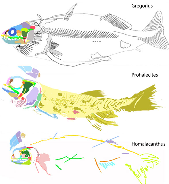
Elopomorphs are not basal bony fish in the LRT where more taxa are included. Prohalecites (Fig. 1) and spiny sharks like Homalacanthus (Fig. 1) are basal taxa to their respective bony fish clades following the first great dichotomy of bony fish. Gregorius (Fig. 1) is their last common ancestor (LCA). Note the shorter rostrum and large eyes close to the anterior margin are juvenile traits retained into adulthood, as we discussed earlier.

Figure 1. Gregorius descends from Hybodus, the shark and is ancestral to Prohalecites at the base of the ray-fin bony fish. Gregorius is also ancestral to Homalacanthus at the base of the spiny sharks leading to lobefins, placoderms, catfish and a variety of other taxa.

Figure 1. Gregorius descends from Hybodus, the shark and is ancestral to Prohalecites at the base of the ray-fin bony fish. Gregorius is also ancestral to Homalacanthus at the base of the spiny sharks leading to lobefins, placoderms, catfish and a variety of other taxa. See figure 2 for a to scale view.

Figure 2. Representatives from the Early Devonian radiation that gave us bony fish, including Prohalecites and Homalcanthus.

Figure 2. Representatives from the Early Devonian radiation that gave us bony fish, including Prohalecites and Homalcanthus. Harpagofututor is close to living moray eels.

A short face and large orbit
characterize both branches of basal bony fish in the LRT, derived from Gregorius, a late survivor ion  the Late Carboniferous of an Late Silurian radiation.

Figure 1. Taxa from the LRT on one branch of the bony fish. Doliodus is one of these.

Figure 3. Taxa from the LRT on one branch of the bony fish. Doliodus is one of these.

Figure 1. Click to enlarge. Acanthodians and their spiny and non-spiny relatives in the LRT (subset Fig. 2), not to scale.

Figure 4. Acanthodians and their spiny and non-spiny relatives in the LRT (subset Fig. 2), not to scale.

Due to taxon exclusion,
Arratia 2010 lists the moray eel, Gymnothorax, and the gulper eel, Eurypharynx (Fig. 5), as elopomorphs. By contrast, when more taxa are added, as in the LRT, both nest closer to Gregorius and hybodontid sharks, both basal to the bony fish first dichotomy.

Figure 6. Eurypharynx evolution. This clade split from Gregorius prior to the major split in bony fish.

Figure 5. Eurypharynx evolution. This clade split from Gregorius prior to the major split in bony fish.

Some taxa (e.g  spiny sharks) at basal nodes in the LRT
are not mentioned by Arratia 2010. Some elops relatives (e.g. the swordfish, Xiphias) are not mentioned by Arratia 2010. Osteoglossum (Fig. 6) nests in the other bony fish clade, the one that includes placoderms, catfish, lobe fins and spiny sharks, and not at the base.

Figure 1. The arowana, an Amazon River predator, nests with Late Jurassic Dapedium in the LRT.

Figure 6. The arowana, an Amazon River predator, nests with Late Jurassic Dapedium in the LRT.

After spending several months 
with ninety+ ray fin fish, trying to shuffle and reshuffle them into their evolutionary order in the LRT (correcting hundreds of mistakes along the way) only a relative few LRT characters turn out to be important for lumping and splitting bony fish. And many of these recur as reversals and convergent trait, making the task more difficult.

  1. Orbit close to the tip of the rostrum, in the middle of the skull vs. close to the rear margin. Sometimes a sword can lengthen the rostrum
  2. Sagittal crest or not
  3. Maxilla either with teeth and a butt joint with the premaxilla, or loosely overlapping the premaxilla without teeth, or other variations
  4. Circumorbital bones absent, or present as a gracile ring, or present as large plates extending toward the preopercular, or as a gracile ring extending to the preopercular
  5. Rostral profile convex or cornered or straight or concave
  6. Coracoid short or long or essentially absent
  7. Parietals meet medially or split by an intervening postparietal
  8. Naris separate from the orbit or confluent
  9. Etc., etc.

Earlier we looked at the various radiations of bony fish
from a variety of spiny sharks in their ancestry (Fig. 4). Since then more taxa have been added, especially on the ray-fin clade. Please see the LRT for the latest cladogram.


References
Arratia G 2010. Critical analysis of the impact of fossils on teleostean phylogenies, especially that of basal teleosts. In: Morphology, Phylogeny and Paleobiogeography of Fossil Fishes Elliott DK, Maisey JG, Yu X and Miao D (eds.): pp. 247-274, 15 figs., 6 tabs. Verlag Dr. Friedrich Pfeil, München, Germany – ISBN 978-3-89937-122-2

https://pterosaurheresies.wordpress.com/2020/05/08/an-unexpected-resolution-to-the-spiny-shark-problem/

Didazoon and Vetulicola: Early Cambrian larvacean and echinoderm ancestors

Urochordates and larvaceans are the relatives
we almost never talk about. Here those long, lost relationships are reestablished.

Didazoon haoae (Figs. 1, 2; Shu et al. 2001; ELI 0000197–0000217) was described as a member of the Ventulicolia, an “enigmatic phylum” of Cambrian deuterostomes of otherwise uncertain affinities. It was a simple, barrel-shaped animal with a line of gill openings dotting a large atrium followed by a down-angled tail. There was no head.

From the Diagnosis:
“Bipartite, cuticularized body, anterior segmented region and voluminous mouth, ventral margin ¯attened, widens posteriorly. On either side the anterior bears fve circular structures, in the form of a cowl with posteriorly directed opening and basin-like interior, apparently connected to interior. A prominent constriction separates dorsal region of anterior from posterior section. The latter, composed of seven segments, tapers in both directions, with rounded posterior termination. Internal anatomy includes alimentary canal, possibly voluminous in anterior, and in posterior narrow intestine, straight or occasionally coiled. Dark strand located along ventral side of anterior section, possibly representing endostyle.”

Figure 1. Didazoon in situ and reconstructed. The 'intestine' may be a notochord remnant because it extends to the tail tip, distinct from Metaspringgina (Fig. 2). The tail was down in all larvaceans.

Figure 1. Didazoon in situ and reconstructed. The ‘intestine’ may be a notochord remnant because it extends to the tail tip, distinct from Metaspringgina (Fig. 2). The tail was down in all larvaceans.

Comparisons to MetasprigginaArandaspis and larvacea
(Fig. 2) were not made by Shu et al. 2001. Here the ‘cuticularized body’ is transitional between Branchiostoma (Fig. 2) and extant larvacea (Fig. 2 which share several traits with these vetulicolians.

In all these taxa, the tail hangs down
and the mouth opens slightly ventrally, beneath what was the rostrum in lancelets. Prior reconstructions at Wikipedia have the tail up and the animal upside-down.

Figure 2. Branchiostoma, Metaspriggina, Didazoon, Vetulicola, Arandaspis and Larvacea to scale. Note the presence of an atrial pore/posterior atrial opening in Branchiostoma and the vetulicolians while Metaspriggina and Arandaspis had fish-like gill openings. The tail of larvaceans hangs down.

Figure 2. Branchiostoma, Metaspriggina, Didazoon, Vetulicola, Arandaspis and Larvacea to scale. Note the presence of an atrial pore/posterior atrial opening in Branchiostoma and the vetulicolians while Metaspriggina and Arandaspis had fish-like gill openings. The tail of larvaceans hangs down.

According to Wikipedia,
Vetulicola (Figs. 2, 3) “is the eponymous member of the enigmatic phylum Vetulicolia, which is of uncertain affinities, but may belong to the deuterostomes. Vetulicola cuneata could be up to 9 cm long (shown at only 6cm in Fig. 2).

Earlier we looked at the parallel development of the enlarged gill chamber in craniates and gnathostomes like the manta ray (Manta) and whale shark (Rhinchodon) that also fed on plankton, but on a much larger scale.

Figure 3. Vetulicola in situ and flipped in vivo configuration.

Figure 3. Vetulicola in situ and flipped in vivo configuration. The shape is transitional to both the larvacea (Fig. 2) and to the crinoid (Fig. 5). Note the four-part separation of the mouth parts, like a budding flower.

According to Wikipedia,
“Vetulicola’s taxonomic position is controversial. Vetulicola cuneata was originally assigned to the crustaceans on the assumption that it was a bivalved arthropod like Canadaspis and Waptia, but the lack of legs, the presence of gill slits, and the four plates in the “carapace” were unlike any known arthropod.”

“Shu et al. placed Vetulicola in the new family Vetulicolidae, order Vetulicolida and phylum Vetulicolia, among the deuterostomes. Shu (2003) later argued that the vetulicolians were an early, specialized side-branch of deuterostomes.”

“Dominguez and Jefferies classify Vetulicola as an urochordate, and probably a stem-group appendicularian (= Larvacea, Tunicata). Like a common tunicate larva, the adult Appendicularia have a discrete trunk and tail.”

“In contrast, Butterfield places Vetulicola among the arthropods.”

“The discovery of the related Australian vetulicolian Nesonektris, from the Lower Cambrian Emu Bay Shale of Kangaroo Island, and the reidentification of the “coiled gut” of vetulicolians as being a notochord affirms the identification as an urochordate.”

Figure 5. Lavacea diagram showing the tadpole organism and the house it builds from cellulose and protein. This is a highly derived organism, based on two parts that originated in the Cambrian with Vetulicola.

Figure 4. Lavacea diagram showing the tadpole organism and the house it builds from cellulose and protein. This is a highly derived organism, with origins in the Cambrian with Vetulicola and close to the salp taxa in figure 3,. Here the larvacea retains the swimming muscles and notochord that extends to the tail tip.

According to Wikipedia:
“Larvaceans have greatly improved the efficiency of food intake by producing a test, which contains a complicated arrangement of filters that allow food in the surrounding water to be brought in and concentrated prior to feeding. By regularly beating the tail, the larvacean can generate water currents within its house that allow the concentration of food. The high efficiency of this method allows larvaceans to feed on much smaller nanoplankton than most other filter feeders.”

“The immature animals resemble the tadpole larvae of ascidians, albeit with the addition of developing viscera. Once the trunk is fully developed, the larva undergoes “tail shift”, in which the tail moves from a rearward position to a ventral orientation and twists 90° relative to the trunk. Following tail shift, the larvacean begins secretion of the first house.”

There are no other chordates that shift the tail 90º relative to the trunk and build an oceanic house of cellulose and protein. Nevertheless Vetulicolia is not a new phylum, but a transitional set of taxa linking lancelets to larvacea and to crinoids. Vetulicolians (Figs. 1–3) demonstrate the down-angled tail appeared in the Early Cambrian pointing to an Ediacaran origin for lancelet-like chordates. That means previously unknown, more active, less benthic (= sea floor) niche full of free-swimming chordates was also present in the Ediacaran.

Figure 7. Vetulicola compared to a fossil crinoid. Note the splitting of the mouth parts into separate 'arms' has only just begun here. The crinoid stalk is the segmented 'tail' of Vetulicola.

Figure 5. Vetulicola compared to a fossil crinoid. Note the splitting of the mouth parts into separate ‘arms’ has only just begun here, starting with four. The crinoid stalk is the segmented ‘tail’ of Vetulicola.

When lips become arms… and tails become stems…
A clade of vetulicolians (former lancelets) also developed a down-hanging ‘tail’ (= stem) that ultimately evolved to become a holdfast: the crinoids within the echinoderms. Remember, adult lancelets also plant their tails in the substrate to become sessile plankton feeders. Crinoids are the ancestors of starfish (Fig. 6), a clade that flips, mouth-side down, and concurrently loses the tail (= stem). Not all echinoderms have a pentagonal morphology. The mouth of Vetucolia has four slightly separating mouth parts. The atrium and tail have just a trace of homologous armor, ringed in the case of the tail and stem.

Figure 3. Chordate evolution, changes to Romer 1971 from Peters 1991. Here echinoderms have lost the tail and gills of the free-swimming tunicate larva.

Figure 6. Chordate evolution, changes to Romer 1971 from Peters 1991. Here echinoderms have a stem-like tail with a holdfast, later lost in starfish, similar to the tail and gills of the free-swimming tunicate larva and adult larvaceans.

Garcia-Bellido et al. 2014 concluded
“Phylogenetic analyses resolve a monophyletic Vetulicolia as sister-group to tunicates (Urochordata) within crown Chordata. The hypothesis suggests that a perpetual free-living life cycle was primitive for tunicates. Characters of the common ancestor of Vetulicolia + Tunicata include distinct anterior and posterior body regions – the former being non-fusiform and used for filter feeding and the latter originally segmented – plus a terminal mouth, absence of pharyngeal bars, the notochord restricted to the posterior body region, and the gut extending to the end of the tail.” 

Garcia-Bellido et al. nested tunicates and vetulicolians as sisters to craniates, both derived from lancelets (cephalochordates). That seems to be correct based on the above figures in their tail-down orientation.

Cameron, Garey and Swalla (2000) reported,
“The nesting of the pterobranchs within the enteropneusts dramatically alters our view of the evolution of the chordate body plan and suggests that the ancestral deuterostome more closely resembled a mobile worm-like enteropneust than a sessile colonial pterobranch.”

Actually Peters 1991 (Fig. 6) published that hypothesis earlier… without the use of DNA.

Now we have long-sought evidence in the form of homologous traits that link vetulicolians to chordates (Fig. 2) and to echinoderms (Fig. 5). All have been understood as deuterostomes. The connection between larva was indicated earlier, according to Cameron, Garey and Swalla 2000. Now we have previously overlooked adult homologs to study and compare. This may be a novel hypothesis of interrelationships. If not, please provide a citation so I can promote it here.

Vetulicolians will not enter the LRT.
They diverge from the vertebrate lineage while keeping some basal lancelet traits and feeding patterns. The degree of their divergence indicates an ancient split from lancelets + fish, just as the divergence of starfish indicate a similar ancient split from lancelet deep time ancestors. They no longer resemble lancelets, except for their cirri-lined mouth parts and attendant mucous strands.


References
Aldridge RJ et al. (4 co-authors 2007. The systematics and phylogenetic relationships of vetulicolians. Palaeontology. 50 (1): 131–168.
Cameron CB, Garey JR and Swalla BJ 2000. Evolution of the chordate body plan: New insights from phylogenetic analyses of deuterostome phyla. PNAS 97(9):4469–4474.
Garcia-Bellido DC et al. 2014. A new vetulicolian form Australia and its bearing on the chordate affinities of an enigmatic Cambrian group. BMC Evolutionary Biology 2014, 14:214 http://www.biomedcentral.com/1471-2148/14/214
Peters D 1991. From the Beginning – The story of human evolution. Wm Morrow.
Shu D-G et al. (8 co-authors) 2001. Primitive deuterostomes from the Chengjiang Lagerstätte (Lower Cambrian, China) Nature 414:419–424.

wiki/Didazoon
wiki/Vetulicolia
wiki/Vetulicola
wiki/Larvacea

.https://www.youtube.com/watch?v=ZXCOZ2_blb8

The lancet fish (Alepisaurus): a swordfish with fangs and no sword

According to Wikipedia
The lancet fish (Alepisaurus) is a traditional member of the Aulopiformes, the clade of lizardfish and their allies. Lizardfish, as you might remember, have eyeballs as far anteriorly as possible, the opposite of lancet fish.

Figure 1. Alepisaurus, the lancet fish nests with swordfish and eels in the LRT.

Figure 1. Alepisaurus, the lancet fish nests with swordfish and eels in the LRT.

By contrast,
in the large reptile tree (LRT, 1810+ taxa) the lancet fish nests close to European eels (Anguilla), but closer to swordfish (Xiphias). All three have similar skulls with a straight rostrum and very little (if any) post-orbital region anterior to the massive hyomandibular. Of course, neither eels nor swordfish have anything like those massive fangs found in the lancet fish. All three had a last common ancestor (LCA) distinct from other fish, and each evolved from that LCA their own special way leaving few clues, other than a similar skull, as evidence of their relationship to one another. That’s the way the LRT recovered it.

Figure 1. Extant swordfish (Xiphias) to scale with Eocene swordfish (Blochius).

Figure 2. Extant swordfish (Xiphias) to scale with Eocene swordfish (Blochius), which looks more like Alepisaurus overall.

That’s why we run all taxa through phylogenetic analysis. 
To do otherwise, to cherry-pick one trait or a dozen, is to “Pull a Larry Martin” which we are cautioned (for good reason) never to do. Professor Martin taught us well.

Figure 5. Skull of Anguilla, the European eel, compares well with that of Bavarichthys. Note the loss and reduction of preorbital bones.

Figure 3. Skull of Anguilla, the European eel, compares well with that of Bavarichthys. Note the loss and reduction of preorbital bones.

Alepisaurus ferox
(Lowe 1833, 1835a b, 215cm) is the extant lancetfish. Traditionally considered a aulopiform (= lizard fish. likeTrachinocephalus) here Alepisaurus nests as a swordfish sister without a rostrum sword, but with giant fangs and vestigial pelvic fins. During prey capture the fangs pierce swimming muscles, stopping the struggle. This hermaphrodite taxon can descend more than a mile into deep unlit seas.

Figure 4. Swordfish ontogeny (growth series). Hatchings have teeth, a short bill and an eel-like body still lacing pelvic fins.

Figure 4. Swordfish ontogeny (growth series). Hatchings have teeth, a short bill and an eel-like body still lacing pelvic fins.

It’s worth noting
that swordfish hatchlings have teeth and a long dorsal fin, like lancet fish. So the resemblance is closer in younger swordfish. It’s also worth noting that fossil swordfish precursors, like Blochius (Fig. 4, Eocene; 60-150cm) also had proportions closer to lancet fish.

Fierstine 2006, in his history of billfishes,
mistakenly included the convergent sailfish, Istophorus, and excluded the European eel and the lancet fish. This is what I mean when we talk about “Pulliing a Larry Martin.” It looks right. It feels right. It gets published. However, whenever you cherry-pick taxa, you are less likely to get the big picture you are chasing: an accurate hypothesis of interrelationships that models real evolutionary events. Instead you should  employ lots and lots of taxa. Then the big picture (= the accurate model) will come to you. The cladogram will recover the correct tree topology.


References
Fierstine HL 2006. Fossil history of billfishes (Xiphoidei). Bulletin of Marine Science 79(3):433–453.
Lowe RT 1833. Description of a new genus of acanthopterygian fishes, Alepisaurus ferox. Proc. zool. Soc. Lond ,1:104.
Lowe RT 1835a. Description of a new genus of acanthopterygian fishes, Alepisaurus ferox. Trans. zool. Soc. Lond., 1: 123-128, pl. 19.
Lowe RT 1835b. Additional observations on Alepisaurus ferox. Trans. zool. Soc. Lond., 1: 395-400, pl. 59.

wiki/Aulopiformes
Alepisaurus_ferox