Every so often an insight comes along that makes one go back and change items that insight applies to. Today most placoderm skull bones remain unchanged, but a few receive new tetrapod homolog identities (Fig 1).
A new genesis for the hyomandibula is the subject of today’s blogpost.
According to Wikipedia, “It [the hyomandibula] usually plays a role in suspending the jaws and/or operculum (teleostomi only). It is commonly suggested that in tetrapods (land animals), the hyomandibula evolved into the columella (stapes).
The stapes is one of the three tiny ear bones in the human skull.
Figure 1. Placoderm skulls with DGS colors applied based on tetrapod homologies. Of interest here in Eriptychius (Ordovician) and Entelognathus (Late Silurian) is the large dark green bone complex: the hyomandibular originating as an external = dermal bone and the cyan bone: the jugal. The preoperulum is the light yellow bone. The quadrate is red. The lacrimal is light tan. No maxilla appears yet here, but a premaxilla has its genesis. The postorbiital (amber) is already in two parts.
Here the hyomandibular has its genesis as a large, flat dermal bone in Entelognathus (Fig 1) and other placoderms (Figs 3–5). The broad hyomandibula later assumes the narrow shape, order and size of a gill bar, as in the Middle Devonian shark, Gladbachus (Fig 2).
Placoderms have shark-like gill bars inside the armored thoracic carapace and plastron (Fig 4), dermal bones that disappear in sharks (Fig 2) and bony fish.
Figure 2. Gladbachus, a basal shark, retained Entelognathus tetrapod homologies (Fig 1). Note the elongate jugal (cyan) and hyomandibular (dark green) duplicated in flat dark green as it is repaired to the in vivo position. The lacrimal (tan) still serves as the large bone support for the thin maxilla layer with the origin of lateral teeth.
According to Wikipedia, “In jawless fishes a series of gills opened behind the mouth, and these gills became supported by cartilaginous elements. The first set of these elements surrounded the mouth to form the jaw. There is ample evidence[3] that vertebrate jaws are homologous to the gill arches of jawless fishes. The upper portion of the second embryonic arch supporting the gill became the hyomandibular bone of jawed fishes, which supports the skull and therefore links the jaw to the cranium.”
Figure 1. Late Early Devonian Stensioella undergoes re-scoring and re-nesting closer to Sphryna, the extant hammerhead shark.
This traditional hypothesis is supported by Gladbachus (Fig 2), but Silurian Bianchengichthys, Devonian Homosteus (Fig 4) and Ordovician Eriptychius (Fig 3) bring new insight to hyomandibula genesis.
Entelognathus primordialis (Zhu et al. 2013; Late Ludlow, Late Silurian, 419 mya; IVPP V18620) is a genus of placoderm fish with tiny eyes. It nests at the transition to Chondrichthyes and Osteichthyes. Here skull bones reidentified with their tetrapod homologies. Pre-teeth are pustules and wrinkles on the bone.
Figure 4. Homosteus, a primitive placoderm, shows an earlier evolution of the hyomandibula (dark green. A) and exposes the gill bars inside the atrium, beneath the here missing carapace of the thoracic armor.
Gladbachusadentatus (Heidtke & Krätschmer 2001; Burrow and Turner 2013; Coates et al. 2018; Middle Devonian, est. 54cm) was originally considered an ‘unfamiliar’ basal chondrichthyan close to acanthodians (spiny sharks). Here it nests withLoganellia. This toothless taxon with a wide gape was also a filter feeder, filtering small prey with enormous gill bars. The unique holotype preserves the pelvic area.
Figure 5. The tiny placoderm, Bianchengichthys, is close to the origin of jaws. Note the presence of the hyomandibula (dark green) and jugal (cyan blue) here.
Eriptychius americanus (Walcott 1892, Bryant 1946; Ordovician) is one known from scattered bones here put together for the first time in a tentative reconstruction based on the bauplan of Late Silurian Entelognathus (Fig 1), Late Silurian Bianchengichthys and Early Devonian Stensioella.
References Heidtke UHJ and Krätschmer K 2001.Gladbachus adentatus nov. gen. et sp., ein primitiver Hai aus dem Oberen Givetium (Oberes Mitteldevon) der Bergisch Gladbach – Paffrath-Mulde (Rheinisches Schiefergebirge). Mainzer geowiss. Mitt. 30, 105–122. Walcott CD 1892. Preliminary notes on the discovery of a vertebrate fauna in Silurian (Ordovician) strata. Geological Society of America Bulletin 3:153-172. Zhu M et al. 2016. A Silurian maxillate placoderm illuminates jaw evolution. Science 354.6310 (2016): 334-336.
Short one today as the graphics (Figs 1–3) tell the tale.
Figure 1. The origin of flying fish and opahs from smaller extant zebrafish (Danio) and Late Jurassic Oreochima AMNH 9910. Note the elongation of the locked-down coracoid. As in birds and pterosaurs this elongation signals the advent of flapping for locomotion.
According to Wikipedia flying fish evolved from Triassic Thoracopterus and kin.
By contrast in the large reptile tree (LRT, 2297 taxa) Triassic Thoracopterus is related to threadfins (Polydactylus), sea robins (Prionotus) and flying gurnards (Dactylopterus). These are all bottom dwellers with large pectoral fins that never try to fly and do not have such a deep and narrow coracoid.
Figure 2. Oreochima ellioti plate and counterplate, along with original diagram and DGS tracing.
Exocoetus volitans (Linneaus 1758; up to 30cm, Fig 1 ) is the extant blue flyingfish, here related to a smaller Jurassic relative, Oreochima (Figs 1–3). Exocoetus travels in schools or schoals. Sometimes they exit the water to avoid predators. Juveniles have a relatively shorter torso. Hatchlings are slow-moving and tiny. Distinctly flying fish and their relatives have a jaw joint directly below the orbit. The coracoid is larger than the scapula, anchoring muscles that power the pectoral fins.
Figure 3. Oreochima specimens AMNH 9911 and 9912. DGS colors applied here. Broken pieces of the preopercular are restore here. This specimen has shorter pectoral fins. Living zebrafish (Danio) have gender color differences. Perhaps elongated pectoral fins first evolved as secondary sexual characters, as in birds and pterosaurs, first on males, then on both genders. Is there a better hypothesis?
Oreochima ellioti (Schaeffer 1972, Early Jurassic, Antarctica, 6cm long) was originally considered close to Pholidophorus. The Bean 2021 thesis nested Oreochima close to Wadeichthys. The AMNH 9920 (FF) specimen nests closer to Wadeichthys. Here the AMMH 9910 specimen nests with the flying fish, Exocoetus.
We looked at the several specimens of little Oreochima earlier here in May 2023. The AMNH 9920 (FF)specimen (Bean 2021a) continues to nest with Wadeichthys.
Living zebrafish (Danio, Fig 1) have gender color differences. Perhaps elongated pectoral fins and deep coracoids first evolved in Late Jurassic Oreochima as secondary sexual characters, as in birds and pterosaurs.
In counterpoint, flying fish are silvery, not brightly colored. They are food for larger, fast surface-dwelling fish, like the Mahi-mahi (Coryphaena). Flying fish ‘wings’ may be large, but they are not flapped for thrust, neither in air nor underwater. Instead, while underwater the pectoral fins are kept close to the body to increase streamlining. In the air, their flying consists of short glides, usually as an escape from predators.
On the other hand, brightly colored opahs (Fig 1) swim by flapping. Link to video here.
References Bean LB 2021a. The morphological revisions of freshwater fishes from Late Jurassic – Early Cretaceous sites in Australia and other Gondwanan continents leads to new phylogenetic hypotheses of relationships among stem teleosts. U3336714 Research School of Earth Sciences. A thesis submitted for the degree of Doctor of Philosophy of The Australian National University. Bean LB 2021b. Revision of the Mesozoic freshwater fish clade Archaeomaenidae. Alcheringa: An Australasian Journal of Palaeontology. 45 (2): 217–259. Linnaeus C 1758. Systema naturæ per regna tria naturæ, secundum classes, ordines, genera, species, cum characteribus, differentiis, synonymis, locis. Tomus I. Editio decima, reformata. Olsen PE 1984. The skull and pectoral girdle of the parasemionotid fish Watsonulus eugnathoides from the Early Triassic Sakamena Group of Madagascar, with comments on the relationships of the holostean fishes. Journal of Vertebrate Paleontology 4(3):481–499. Piveteau J 1930. C R Acad. Sci. Paris, 191. Piveteau J 1935. Palkontologie de Madagascar. XXI. Les Poissons du Trias infkrieur. Contribution 2 l’ktude des actinoptkrygiens. Annales de Pal6ontologie 23:8 1-1 80. Schaeffer B 1972. A Jurassic fish from Antarctica. American Museum Novitates 2495:1–17. Günther A 1868. Catalogue of the fishes in the British Museum. Trustees, British Museum, London, 512 pp. http://dx.doi.org/10.5962/bhl.title.8314 Schaeffer B 1972. A Jurassic fish from Antarctica. American Museum Novitates 2495:1–17.
Short one today as graphics (Fig 1) and a short text tell the tale of an overlooked interrelationship, finally explored. If you look at enough fish long enough some eventually start to look alike. This is called the learning process.
Figure 1. Early Silurian Shenacanthus and Late Mississipian Ozarcus share more traits with each other than with other tested taxa. Each one informs the other with regard to missing parts. Shown slightly smaller than life size on 72dpi monitors, plus enlargements of Shenacanthus to show details. That’s a portion of the hyomandibular (dark green) benath the intertemporal (yellow green). The face had to be reassembled and parts identified.
Interrelationships, like this, become more obvious after taxa are put together for the first time. Otherwise, as in this case, almost a year can go by without having that eureka moment, like recognizing twins for the first time in a large high school. Both taxa were always close to one another in the LRT near the dichotomy of sharks and bony fish following tiny derived placoderms in the Early Silurian. Now they are closer.
Shenacanthus entered the LRT almost a year ago, October 1, 2022. Ozarcus was always an oddity among the sharks.
Recognizing the (red) quadrate on Ozarcus was not a labial cartilage was helpful today. The multiple gill bars and claspers are traits shared with sharks.
The missing thoracic shield on Ozarcus is a trait of most descendants of placoderms, including bony fish, sharks and humans. Some spiny sharks close to Shenacanthus retain a thin and reduced version of this shield set. A much modified and much later reappearance of a carapace and plastron in various armored taxa (turtles, placoderms, ankylosaurs, glyptodonts, etc.) is an intriguing reversal hypothesis, now that we know placoderms were our ancestors. So, it remains in our genes.
An Early Silurian split between sharks and spiny sharks (= basal bony fish) is documented by these taxa.
Shenacanthus vermiformis (Zhu et al 2022, Early Silurian, est 3cm long) is the earliest known of the jawed placoderms and the basalmost member of the spiny sharks (Osteichthys). That makes it very close to basal sharks. Claspers are present. Ozarcus is a much larger and much later sister from the Late Mississippian.
Ozarcus mapesae (Pradel et al. 2019; Early Carboniferous, 325 mya; AMNH FF20544) nests with the much smaller basal osteichthyan, Shenacanthus, in the LRT. Pradel et al 2014 considered the palatoquadrate (pq) a single bone rather than the lacrimal shown here. The intertemporal anchors the hyomandibular here (eh) as in other vertebrates. The teeth are extremely tiny, much smaller than each tooth basin in the maxilla. This appears to be a derived trait, because Shenacanthus has relatively larger maxillary teeth. The premaxilla is a tiny toothless area between the incurrent + excurrent nares. The seemingly crushed narrowness of the skull is odd, perhaps unique. The thoracic shield of its ancestor was not preserved here, but is preserved in Mesacanthus and similar relatives.
Apologies for late replies to comments. Housekeeping progress (like this) continues in the fish subset of the LRT, which includes some 485 taxa. Each score and suture for each taxon is under review, so focusing on the few remaining problems with earnest turns them into solutions and possible hypotheses a little more quickly.
References Pradel A, Maisey JG, Tafforeau P, Mapes RH and Mallant J 2014. A Palaeozoic shark with osteichthyan-like branchial arches. Nature 13185. doi:10.1038/nature13195e Zhu Y-A et al (10 co-authors) 2022. The oldest complete jawed vertebrates from the early Silurian of China. Nature 609:954–958. online
The authors missed the point of their new fossil find. They found a bipedal protorosaur (Fig 1), the first obligate biped from that clade. And it was found in the same bed as Ixalherpeton(Fig 1), another likely bipedal protorosaur.
Müller et al 2023 described a Late Triassic partial skeleton (CAPPA/UFSM 0356) as a lagerpetid, consisting of an incomplete skull, some vertebrae, a partial pelvis and limb elements (Fig 1). Venetoraptor gassenae is estimated at nearly a meter long and a third of a meter tall at the hips. Müller et al, like others before them, are spreading the word that lagerpetids were close to the origin of dinosaurs and pterosaurs, following the current myth taught in university textbooks.
I’d like to see the pes to see if Venetoraptor walked on two toes (3 and 4), as in Lagerpeton. If you have a jpeg of the Venetoraptor pes, please send it. Only toe 3 was shown in the paper (Fig 1).
Figure 1. Venetoraptor compared to the protorosaur, Ixalerepton, and two Triassic fenestrasaurs, Cosesaurus and Bergamodactylus to scale. Müller et al argue for a relationship between these taxa. Seems odd based on size alone. One only needs to add taxa to figure this out for oneself. The manus of Prolacerta also has a longer mc4, a trait not seen in later archosauriformes.
From the abstract: “Its skull has a sharp, raptorial-like beak, preceding that of dinosaurs by around 80 million years, and a large hand with long, trenchant claws that firmly establishes the loss of obligatory quadrupedalism in these precursor lineages. Combining anatomical information of the new species with other dinosaur and pterosaur precursors shows that morphological disparity of precursors resembles that of Triassic pterosaurs and exceeds that of Triassic dinosaur.”
That sharp beak (premaxilla) and large trenchant claws are presently unique to Venetoraptor among the Protorosauria. Those bones were not preserved in Ixalerpeton (Fig 1), which may also have been a facultative biped based on proportions.
Figure 2. Above: Venetoraptor from Muller et al 2023. Colors added here. At bottom Prolacerta has a similar skull. The hooked premaxilla (yellow) of Venetoraptor has a homolog in the downturned premaxilla of Prolacerta.
When added to the large reptile tree (LRT, 2297 taxa) Venetoraptor nests within the clade Protorosauria. Like Prolacerta (Fig 3), the tibia + fibula is longer than the femur, the chevrons are deep and metacarpal 4 is slightly longer than mc3. Also, look for a sliver of a supratemporal. By contrast, in archosaurs mc4 is usually shorter to much shorter than mc3.
The manus remains unknown in lagerpetids, going back to Diandongosuchus, an outgroup taxon to the Proterochampsidae, the clade that includes Lagerpeton and Tropidosuchusin the LRT. These two taxa walk on pedal digits 3 and 4, distinct from all other vertebrates sans Deinonychus and kin.
By contrast, you’ll recognize pterosaurs and their ancestors by their attenuated tail, elongate ilia framing 4+ sacrals, prepubes, four toes pointing anteriorly and toe five usually flexed under to point posteriorly impressing the toe knuckle in Rotodactylus tracks
Unfortunately,Venetoraptor scores too few characters to remain in the LRT, which minimizes taxon exclusion by including a large number and wide gamut of largely complete taxa that result in very few nodes where resolution is lost.
Figure 3. Ixalerpeton pelvis compared to Prolacerta.
If you want to learn more about pterosaur ancestry, click here, here, here,here, or here (at RearchGate.net, also see Fig 4). A timeline of pterosaur origin studies can be found here. You’ll note a two-decade pattern of taxon exclusion has hobbled pterosaur origin studies. That continues in Müller et al 2023. Apparently no one wants to study the pertinent complete fossils with soft-tissue preservation, preferring instead to employ other taxa that share very few traits with pterosaurs.
Thank you to reader UE who raised the issue of Venetoraptor.
Figure 4. Click to enlarge. The origin of the pterosaur wing and the migration of the pteroid and preaxial carpal. A. Sphenodon. B. Huehuecuetzpalli. C. Cosesaurus. D. Sharovipteryx. E. Longisquama. F-H. The Milan specimen MPUM 6009, a basal pterosaur.
PS The last week of no posts and no replies to comments were due to concentrated study on fish. Many mistakes were corrected. Hopefully all freshman naiveté issues will be resolved soon. The allometric growth patterns of fish, the loss and gain of facial bones during neotony, convergent finless taxa,?convergent catfish-like taxa and just recognizing overlooked homologies has been difficult, but filled with eureka moments. You’ll see the results in future posts.
PPS Only one toe (digit 3, Fig 1 was illustrated in this paper, which is why I asked if the foot was available.
The Müller et al cladogram suffered from massive taxon exclusion. It included some lepidosaurs, but did not include the basal tritosaur, Huehuecuetzpalli. Nor did the authors cladogram find a basal split between lepidosauromorphs and archosauromorphs, which is only recovered when you include Early Carboniferous Silvanerpeton in your own LRT. Cosesaurus (Fig 1), Sharovipteryx, Longisquama and Bergamodactylus (Fig 1) were not included. These taxa are transitional to pterosaurs in the LRT (and Peters 2000). Tropidosuchuswas included. It attracts Lagerpeton to the Proterochampsidae in the LRT, but not in Müller et al. The authors nested the thalattosaur, Vancleavea, within the Proterochampsidae because no other thalattosaurs were tested.
Build your own LRT so your paper won’t suffer from this sort of taxon exclusion.
References Müller RT et al (8 co-authors) 2023. New reptile shows dinosaurs and pterosaurs evolved among diverse precursors. Nature. 620 (7974): 589–594. Peters D 2000b. A Redescription of Four Prolacertiform Genera and Implications for Pterosaur Phylogenesis. Rivista Italiana di Paleontologia e Stratigrafia 106 (3): 293–336. Peters D 2009. A reinterpretation of pteroid articulation in pterosaurs. Journal of Vertebrate Paleontology 29: 1327-1330. Peters D unpublished. Cosesaurus aviceps, Sharovipteryx mirabilis and Longisquama insignis reinterpreted. ResearchGate.net
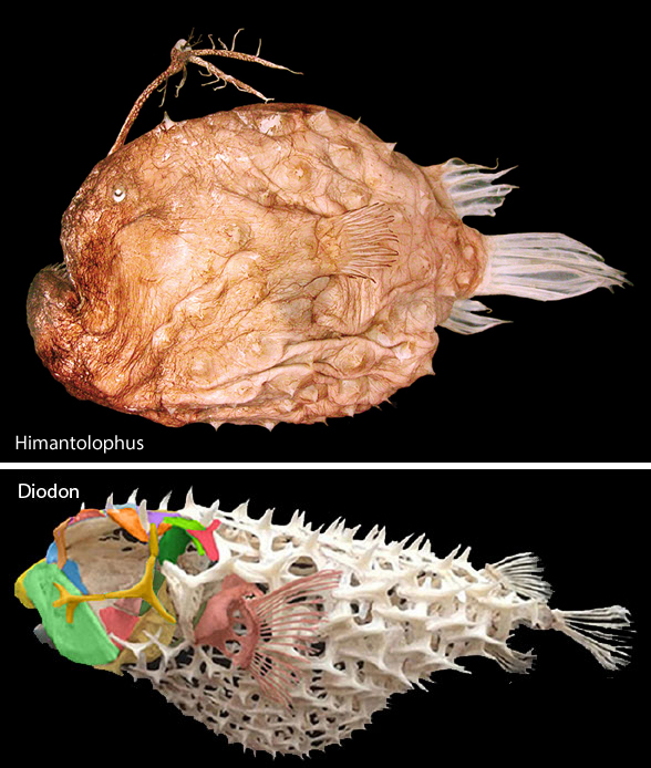
Short one today. More housekeeping in the fish subsection of the large reptile tree (LRT, 2297 taxa) is driving taxa together that have not been nested together before. Today the football fish,Himantolophus, nests with the porcupine fish, Diodon (Fig 1) in the LRT.
Himantolophus is a traditional anglerfish. In the LRT it is now convergent with anglerfish, which are closer to scorpionfish and perch.
Seems obvious now, especially noting that Himantolophus also lacks pelvic fins and has dermal spines all over its body. This hypothesis of interrelationships could change tomorrow. And that’s OK.
Himantolophus groenlandicus (Reinhardt JCH 183; 60cm in length) is a deep sea ‘angler’. Strongly resembling the frogfish Antennarius, Himantolophus lacks any trace of a pelvis or pelvic fin. In the LRT it is more closely related to Diodon the porcupine fish (Fig 1). The skin is covered with large bony plates, each with a medial spine. The teeth are depressible. This single genus has 18 species.
BTW, the LRT has not been updated for the past week and cannot be updated for awhile considering the current wave of changes, like this one (Fig 1), coming into the fish subset of the LRT.
Thank you for your readership, yes, even you certain readers, who hate DGS, the LRT and every time I post here. I realize this is frustrating for you.
Solution: Build your own LRT. Then you’ll have evidence to counter hypotheses made here and elsewhere. Become a player. It’s more fun than sitting in the stands.
Long time readers have been following this 12-year phylogenetic experiment in real time, including mistakes, discoveries, corrections and new hypotheses of interrelationships. There has been only one reason to build the LRT: it has never been done before, not with this wide gamut of 2297 taxa (Chordates). This analysis needed to be done because taxon exclusion is the number one problem in vertebrate paleontology.
There is no need to cite hypotheses presented here. Just add pertinent taxa to your own more focused studies.
References Reinhardt JCH 1837. Ichthyologiske bidrag til den Grönlandske fauna. Kgl. Danske Vidensk. Selsk. Natur. Math. Afhandl., 7: 83-196, pls. 1-8.
Today we return to a controversy that began a few days ago when the vertebral series of Perucetus (Figs 1–4) was introduced to the world. Many videos and articles followed this announcement.
Science writer Flora Graham wrote an editorial for Nature on Perucetus Here’s the subhead with a modern tie-in. “Perucetus colossus, an ancient whale that lived 38 million years ago, is a new contender for the heaviest animal ever. Plus, four key questions on the new wave of anti-obesity drugs.”
I’ve never seen Nature add a commercial to their subheads before. Funny that they connected “anti-obesity drugs” to the “heaviest animal ever.”
Figure 1. Comparing an average Balaenoptera and the only Perucetus lumbar vertebrae. The extant mysticete has a much larger centrum cross-section. The extinct taxon has much more robust transverse processes, adding to the weight of the bone, likely for bottom-feeding in the clam beds it was found in. Note the blue whale vertebrae is shown anterior to the right, reversed from the Perucetus vertebrae, anterior to left.
Getting back to science. Perucetus and its authors (Bianucci et al 2023) have more than a few followers. Among them is science writer, Flora Graham, in an editorial for Nature. Her headline, “Giant ancient whale could be heavier than blue whale” is ambiguous, but juicy. Graham didn’t say ‘heavier than the largest ever blue whale’, which measured 110ft or 33.5m. The average size for the blue whale is 80 to 90 feet long, shorter for males.
So there’s lots of wiggle room here to boost that Perucetus headline.
Figure 2. How to estimate the size of something if adult human males are your only yardstick. I’m tall so my ulna is 29cm long. The average male has a shorter ulna: 27.4cm..
Facts and estimates Weight of largest ever blue whale: 418,878 pounds or 209 tons. What Graham wrote for Nature:“Most blue whales weigh around 100–150 tonnes; the researchers’ best guess is that Percetus colossus weighed around 180 tonnes.” Wikipedia reports for Perucetus, “weight ranging from 85–340 t (84–335 long tons; 94–375 short tons),”
So there’s lots of wiggle room here, too, especially for a set of Perucetus lumbars (Fig 3) that are not larger than those of an average blue whale (Fig 4). Bianucci et al recognized these estimates were wide-ranging, to their credit.
Figure 3. Comparing three different scale bars vs the Perucetus vertebrae. The longest possible measurement of the known bones of Perucetus based on published scale bars indicates a length of 4.6 meters for the set. So published scale bars were within the realm of correct. Measuring with human yardsticks (Fig 2) gives a shorter value for the lumbar series. Note the neural spine orientation changes in this series, distinct from other whales (Fig 4).
Environomental writer, Emma Marris, also writing for Nature had this to say: “might have been heavier than a blue whale, even if it was not as long.” “The whale’s bones were so enlarged that they look swollen, almost like inflated balloons.” “The bones lacked the porous, spongy structure of typical bones.”
The last two are true. The first is a big maybe not based on current evidence (Fig 4).
Figure 4. Perucetus compared to Basilosaurus, Balaenoptera and Cynthiacetus to scale. As shown in figure 3, the last two vertebrae tapered toward the tail. So reconstructions that show a longer line of lumbars, as in basiolosaurids, do not appear to fit the bones. Short neural spines are found in Desmostylus (Fig 7). Perucetus has a transitional length of neural spines. Perucetus had shorter thoracic ribs than Balaenoptera.
Bone density is related to buoyancy. Densely-boned animals tends to sink. Whales of all types, despite their less dense vertebrae, generally sink when dead, providing islands of nutrition on the sea floor for years thereafter. By contrast, right whales tend to float, at least at first, which made them the ‘right’ whales to harpoon back in the day. Hippos are able to walk on river floors at least partly due to denser than typical limb bones. The marine hippos, the desmostylians, had a presumed similar lifestyle grazing the shallow sea floor, even though they had whale-like bone density in their vertebrae (Hayashi et al. 2013).
If Perucetus grazed the sea floor aided by heavy bone density, and, at best, vestigial hind limbs, we might assume it lived and fed in waters no deeper than its nostril height – which means rather shallow waters. Perhaps other factors enable it to float over deeper waters.
Interesting parameters.“Although manatees are excellent swimmers, the deepest an individual has been seen diving is 10 m (33 ft.). They normally feed no deeper than about 3 m (10 ft.) below the surface of the water.
Why did Bianucci et al focus on Late Eocene Cynthiacetus peruvianus (Fig 4) for their Perucetus Bauplan? Both are from Peru and both have similar morphologies if one simply expands each vertebra in Cynthiacetus to match Perucetus. Why did Bianucci et al not consider a desmostylian or mysticete connection? Probably because all the published literature ignores desmostylians as candidate ancestors for mysticetes. Most of the published literature considers archaeocetes basal to all extant whales.
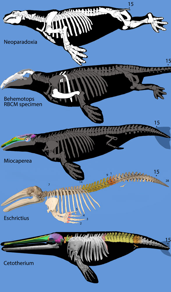
Figure 5. Rorqual evolution from desmostylians, Neoparadoxia, the RBCM specimen of Behemotops, Miocaperea, Eschrichtius and Cetotherium, not to scale. Note the tightly packed lumbars of Cetoterhium, much like Perucetus. Dark gray bones are hypothetical based on phylogenetic bracketing.
Short pre- and post-zygopophyses on Perucetus (Fig 3) would appear to indicate a short range of motion. The brevity of the transverse processes is a second indicator. The brevity of the neural spines is a third indicator. Given these parameters, perhaps the tail of Perucetus was not the massive organ of propulsion seen in basilosaurids and extant whales that have taller, longer, narrower, lighter vertebral processes (Fig 4). Given these parameters the authors considered Perucetus to be a slow moving marine mammal, like a manatee. Along the same lines, let’s not omit the possibility of a very large desmostylian transitional to its own lineage of mysticetes (Fig 5) based on that massive lumbar series and tiny pelvis (Fig 7).
The Barnucci et al SuppData indicated greater flexibility in the lumbar region: 26º dorsal, 60º indicated ventral – but 160º demonstrated ventral, and 17º lateral flexion.
Distinct from other whales of all types, the Perucetus neural spines did not all lean the same way (Fig 3). They diverged in the front and back. In other mysticetes, Ziphius and Orcinus, all the neural spines lean posteriorly. In terrestrial mammals, Delphinus and deep-diving Physeter, the sperm whale, the neural spine angles converge over the anterior caudals.
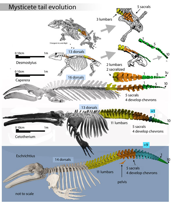
Figure 6. The origin of a robust tail in the ancestor of mysticetes (baleen whales). Note the paddle-like ribs on Caperea and Cetotherium, like those found with Perucetus (figure 4). Note the short, low transverse processes on the long lumbar region of Cetotherium and Eschrichtius AND Perucetus – but not the same in Caperea, which has a short lumbar region and more elongate centra.
Miocaperea (Fig 5 middle) is a Miocene mysticete or pre-mysticete from Peru. Too bad it is only known from a skull. We can only speculate on the post-cranial anatomy. Too bad Eocene Perucetus is only known from vertebrae. We can only speculate on the cranial anatomy. Both are considered bottom-feeders in shallow waters based on the phylogenetic proximity of Miocapera to desmostylians and the extreme density of the Perucetus vertebrae. Let’s keep open the possibility that these two taxa might be related to one another.
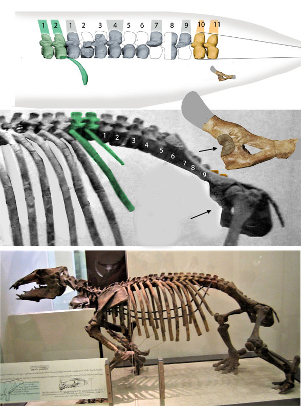
Figure 7. Desmostylus skeleton compared to Perucetus lumbars and pelvis, not to scale. The short neural spines are similar to Perucetus, but the high transverse processes are more primitive in Desmostylus. The pelvis shapes are similar, but the sizes are not. Note the acetabulum contacts the anterior margin in both taxa. The LRT nests desmostylians basal to mysticetes.
Here’s an interesting fact with phylogenetic significance. “Eight months after conception, at the height of the wet season, female hippos give birth to one calf at a time, either on land or underwater. Afterwards, mothers leave the herd for a short period of time to bond with their calves underwater. After a few weeks, the calves finally exit the water to feed on grass.”
Since mysticetes give birth only underwater, we can assume transitional taxa, like desmostylians, moved increasingly to giving birth underwater.
Miocaperea pulchra (Bisconti 2012; late Miocene, 7–8 Ma; 40cm skull length) was considered a Miocene ancestor of Caperea. Here it nests between Behemotops (Fig 5) and Eschrichtius, the gray whale (Fig 5). The post-crania in Miocaperea is not known, so it could have had legs or flippers.
YouTube video in Spanish. Turn on Closed Caption (cc) and translate to English or your favorite language in YouTube settings.
The above 1:11 hour YouTube video is informative, if not a little rambling, joking and opinionated. To his credit, the narrator emphasizes all numbers are estimates. To his credit the narrator realizes the environment must have been rich in food for Perucetus to grow so large.
References Bianucci G et al (15 co-authors) 2023. A heavyweight early whale pushes the boundaries of vertebrate morphology. Nature. doi:10.1038/s41586-023-06381-1 Hayashi S et al (8 co-authors) 2013.Bone Inner Structure Suggests Increasing Aquatic Adaptations in Desmostylia (Mammalia, Afrotheria). PlosOne https://doi.org/10.1371/journal.pone.0059146 Graham F 2023. Daily briefing: Giant ancient whale could be heavier than blue whale. Nature Daily Briefing Online here. Marris E 2023. Could this ancient whale be the heaviest animal ever? Nature News Online here. Peters D unpublished. The triple origin of whales. ResearchGate.net georgiasfossils.com/12c-cynthiacetus-revised
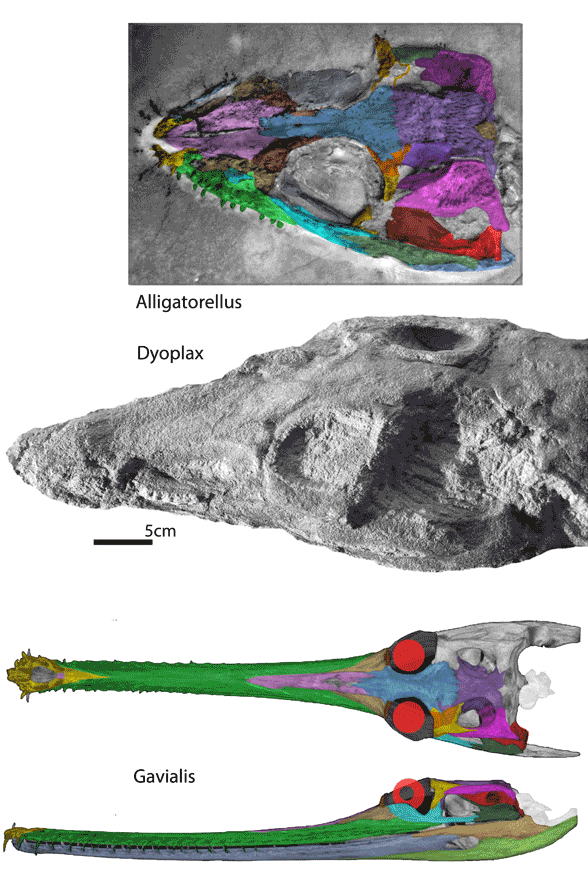
Just another experiment trying to figure things out. Here the Early Triassic crocodylomorph, Dyoplax, was traced again using DGS methods. Then missing and disturbed parts were restored (Fig 1) based on sister taxa in the large reptile tree (LRT, 2297 taxa, subset Fig 2). Those sisters are (at present) Late Jurassic Alligatorellus (Fig 1) and extant Gavialis (Fig 1).
Figure 1. Alligatorellus, Dyoplax and Gavialis compared. Missing and disturbed parts of Dyoplax are restored here.
Dyoplaxarenaceus (Fraas 1867, Lucas, Wild and Hunt 1998; Maisch, Matzke and Rathgeber 2013) is a unique Late Triassic crocodylomorph, one of the three original crocodylomorphs in the 19th century (along with two aetosaurs). Dyoplax nests in the LRT as a basal gavialid and close to Jurassic sea crocodiles.
Alligatorellus beaumonti (Gervais 1871; Late Jurassic) was found in Solnhofen marine limestones. It is a small, short-snouted primitive carnivore, closely related to Late Triassic Dyoplax and the gharial, Gavialis. It has juvenile proportions, so could represent a neotonous adult.
Figure 3. Subset of the LRT focusing on Crocodylomorphain 2023. The new nestings represent just the latest of a changing hypothesis of interrelationships.
A paper on Gavialis phylogeny (Gold, Brochu and Norell 2014) reported, “Some areas of the tree remain controversial, particularly near the base of the crocodylian evolutionary tree. Among these is the phylogenetic position of the Indian gharial, Gavialis gangeticus, whose phylogenetic placement changes dramatically depending on which types of data are analyzed.”
The authors used phylogenetic analysis and genomic analysis. They reported, “The braincase data alone produced a clade that included crocodylids and Gavialis,whereas the Eustachian data resulted in Gavialis being considered a basally divergent lineage.” The authors concluded, “In this study, when the morphometric data were added to discrete morphological and molecular characters, they each result in the identical topology in which Gavialis and Tomistoma combined are closest relatives within Crocodylidae.”
By contrast, in the LRT (Fig 3) using trait analysis Tomistoma (the false gharial) nests basal to Alligator and Crocodylus, apart from Gavialis (the gharial).
Data is not always complete pristine and incontrovertible. Even so, use the data you have and keep looking for improvements, even if they upset / modify your current hypothesis. Run the numbers in your own LRT to find out for yourself.
PS. Croc expert and paleontologist Christopher Brochu questioned the colored division of the traditional croc postorbital into postorbital (yellow) and postfrontal (orange) regions in figure 1 above. He wrote, “Crocodylians don’t have postfrontals at any point in development. They’re not fused to the postorbital – they’re absent entirely. And even if the two bones were fused (which they’re not, since one of the two bones never appears), I have no idea how you would distinguish them on an actual skull. I would recommend re-coloring the postorbital as a single bone, because that’s what it is.”
A closer look at the bones should be educational. My reply and request follow.
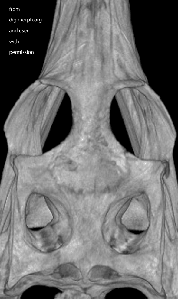
Figure X. Dorsal view of a Gavialis skull from Digimorph.org. Frame 2 boosts contrast. Frame 3 adds colors to indicate where I see a postfrontal (orange) on top of the postorbital (yellow). The left side appears to have suffered a bit of damage (missing part of the frontal to expose the underlying parietal in lavender).
Thanks, Chris, I appreciate your extensive experience with crocodilians. To your point, the vast majority of derived crocodylomorphs have obliterated the suture between the postorbital and postfrontal. To call the remaining bone a postorbital is appropriate because the postfrontal appears to phylogenetically shrink relative to the postorbital.
The LRT nests Gavialis apart from crocs + gators, so Gavialis might not follow crocodilian rules. Brochu 1997 also split Gavialis apart from other crocodilians. That paper did not include Alligatorellus and Dyoplax (Fig 1). The LRT nests Gavialis with these hatchling-sized adult taxa that appear to have a suture between the postorbital and postfrontal. As always, it is hard to tell.
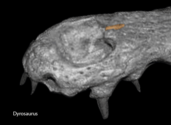
A few crocodylomorphs appear to keep the suture between the postfrontal and postorbital. Dyrosaurus (Fig y) related to Gavialis in the LRT, Fig 2) appears to be one.
Figure Y. Dyrosaurus appears to keep a separation between the postfrontal and postorbital in this adult specimen. It is related to Gavialis in the LRT.
Can you provide evidence of a postorbital-postfrontal complete fusion (obliterated suture) in a gharial that matches the seamless condition in crocs and gators?
While waiting for your evidence, I posted an image from Digimorph.org of a Gavialis skull in dorsal view (Fig x). Frame 2 boosts the contrast. Frame 3 colorizes what little remains of the postfrontal largely atop the postorbital. The bones are fused. Faint outlines appear to remain. There appears to be some damage on the left.
Rather than posting a comment on this image, it would be better if you repeated this tracing exercise and send the results for posting here,. That way we will have two players on the field, rather than one player and one umpire.
I sought images of a Gavialis hatchling skull, but none were found.
PPS – a day later Chris Brochu replied, “If you can demonstrate that any of the points I’ve made is inaccurate, please let me know.”
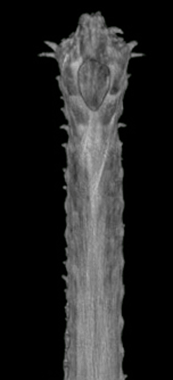
Well here’s the snout of Gavialis. This is the demonstration Brochu asked for. Frame 1 shows the Digimorph.org image alone. Frame 2 shows the snout with the premaxillae (yellow) blocking the nasals from contacting the naris, which is not seen in most tetrapods, including other crocodylomorphs, but is seen in certain extinct crocodylomorphs. Frame 3 shows the nasals (pink), if present, contacting the naris. If the nasals do not contact the naris, at least readers can see the reason why this appeared to be so from this image. See other related taxa shown below ((Fig YY, ZZ) for relevant homologies.
Figure XX. Dorsal view of Gavialis snout. Frame 2 shows the premaxillae blocking nasal contact. Frame 3 shows nasal (pink) contact, as in figures YY and ZZ. This is a younger specimen than the one shown below in figure x.
Figure YY. Snout diagram of Isisfordia showing the nasals contacting the naris.
Figure ZZ. Snout of Dyrosaurus showing the nasals contacting the naris in the manner of Gavialis.
At some point a judgement can be rendered. Are postfrontals present or not (Fig X)? Does the nasal contact the the naris or not (Fig XX)? Sometimes the answer is clear (Figs YY and ZZ). Sometimes the answer is not so clear (Fig XX).
This question is a microcosm of the DGS method. DGS can demonstrate observations to readers. On the other hand, when a scientist reprorts, “Having seen the specimen you’re colorizing… I can assure you that your sutural reconstructions are inaccurate” does not demonstrate observations to readers. It must be believed. No evidence or demonstration is necessary. Statements like this are appeals to authority. They should not be questioned, even with evidence.
For many people, appeals to authority are attractive. That’s why we still have bat-wing birds. Some readers will latch on to Brochu’s appeal to authority in order to blackwash the entire LRT and the DGS method. That is regrettable, but that happens.
I prefer to present evidence, however tenuous (Fig 1). I agree, the evidence for the nasals contacting the naris in Gavialis is tenuous, at present, depending on shadows and light and homologies in closely related taxa. A µCT scan should be able to determine whether of not the nasals beneath the maxillae and premaxilla still contact the naris – or not. It might vary among individuals. So there’s a solution to this argument. Don’t make this a tempest in a teapot. We can figure this out, or let it drop.
It’s not life-or-death. It’s an idea.
FIgure x. Added more than a week later following a citation to this figure, which purports to show the truncation of the nasals in Gavialis. Note the nasals extend to the naris in the other crocodilians. Note also the tiny medial bone (pink arrow) in the top illlustration of the Gavialis snout in the exact place occupied by the nasals in other crocs. By this evidence the middle portion of the Gavialis nasal has been lost, leaving only a small anterior portion (extent unknown below the overlapping premaxilla) and a larger posterior portion, now separated from the anterior. Excellent use of DGS, by the way. Glad to see other workers adopting this technique for demonstration and teaching.
References Fraas O 1867.Dyoplax arenaceus, ein neuer Stuttgarter Keuper-Saurier. Jh. Verein vaterländ. Naturk. Württemberg 23:108-112; Stuttgart. Gold MEL, Brochu CA and Norell MA 2014. An Expanded Combined Evidence Approach to the Gavialis Problem Using Geometric Morphometric Data from Crocodylian Braincases and Eustachian Systems. PLoS ONE 9(9): e105793. doi:10.1371/journal.pone.0105793. Lucas SG, Wild R, Hunt AP 1998.Dyoplax O. Fraas, a Triassic sphenosuchian from Germany. Stuttgarter Beiträge zur Naturkunde, B. 263: 1–13. Maisch MW, Matzke AT and Rathgeber T 2013. Re-evaluation of the enigmatic archosaur Dyoplax arenaceus O. Fraas, 1867 from the Schilfsandstein (Stuttgart Formation, lower Carnian, Upper Triassic) of Stuttgart, Germany. Neues Jahrbuch für Geologie und Paläontologie – Abhandlungen. 267 (3): 353–362.
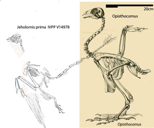
From the abstract “We provide direct dietary evidence that Jeholornis consumed leaves likely from the magnoliid angiosperm clade, and these results lend further support for early ecological connections among the earliest birds and angiosperms. Morphometric reanalysis of the lower jaw of Jeholornis further supports a generalized morphology shared with other herbivorous birds, including an extant avian folivore, the hoatzin.”
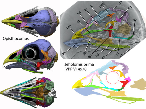
Indeed, the skull of Ophisthocormus, the hoatzin (Figs 1, 2), does greatly resemble (by convergence) that of the new Cretaceous plant-eater, Jeholornis (Figs 1, 2).
Figure 1. Jeholornis and Opisthocomus both ate plants by convergence.
The question is is the new Jeholornis prima conspecific with the holotype (Fig 3)? The holotype has teeth, which are lacking in the new specimen. A distinct skull shape is also notable.
Figure 2. Opisthocomus skull in 3 views compared to the skull of Jeholornis in situ and reconstructed using DGS methods.
Figure 3. Jeholornis prima holotype compared to the new plant-eating specimen. Are they conspecific? Apparently no, but close enough to nest together in the LRT.
When added to the large reptile tree (LRT, 2299 taxa), the three Jeholornis specimens nest together. Their differences are not so great as to separate them = moving them closer to other taxa.
References Yan Wu, Yong Ge, Han Hu, Thomas A. Stidham, Zhiheng Li, Alida M. Bailleul and Zhonghe Zhou, 2023. Intra-gastric phytoliths provide evidence for folivory in basal avialans of the Early Cretaceous Jehol Biota by 28 July 2023, Nature Communications. DOI: 10.1038/s41467-023-40311-z
Publicity from SciTechDaily.com “New findings from the analysis of a 120-million-year-old fossil skeleton of the extinct bird Jeholornis, unearthed from northeastern China, present the earliest known evidence of leaf-eating birds, marking the earliest known evolution of arboreal plant-eating among birds.
“The pheasant-sized Jeholornis, a member of the second most primitive lineage of known birds, has teeth and a long bony tail like its predatory, feathered dinosaur relatives. However, microscopic analysis of the fossilized residues in the stomach of this juvenile, arboreal (tree-living) bird demonstrates that Jeholornis was not a predator. It had eaten tree leaves from a group of flowering plants called magnoliids that includes the living magnolia, cinnamon, and avocado trees.
“After comparison with over 4,000 kinds of modern phytoliths, we can see that most of the identifiable fossil phytoliths from the stomach come from the leaves of magnoliids,” said Dr. Wu Yan from IVPP, first author of the study.
“To further support their hypothesis of leaf-eating in this early bird, the paleontologists also compared the lower jaw of this bird to living birds with a wide range of diets. Coauthor Dr. Hu Han from Oxford University said, “A more detailed statistical analysis of the three-dimensional shape of the lower jaw of Jeholornis shows similarities to the shapes of living birds that eat mostly plants including the living leaf specialist, the hoatzin from tropical forests in South America.”
From the Fang et al abstract: “Modern baleen whales are unique as large-sized filter feeders, but their roles were replicated much earlier by diverse marine reptiles of the Mesozoic.
Replicated? Which taxa? If you can’t come up with a short list, readers will consider this sentence fluff = pushing a narrative lacking evidence or a list to check. Provide the evidence. Provide the list of diverse marine reptile filter feeders.
“Here, we investigate convergence in skull morphology between modern baleen whales and one of the earliest marine reptiles, the basal ichthyosauromorph Hupehsuchus nanchangensis, from the Early Triassic, a time of rapid recovery of life following profound mass extinction.
“Two new specimens reveal the skull morphology especially in dorsal view.”
“The snout of Hupehsuchus is highly convergent with modern baleen whales, as shown in a morphometric analysis including 130 modern aquatic amniotes.”
Notable by their absence: any taxa from the clade Mesosauria, which actually have baleen-like procumbent needle-teeth and phylogenetically precede hupehsuchids in the large reptile tree (LRT, 2297 taxa).
Build your own LRT to find this out for yourself.
“Convergences in the snout include the unfused upper jaw, specialized intermediate space in the divided premaxilla and grooves around the labial margin.’
Maybe not (See Fig 1).
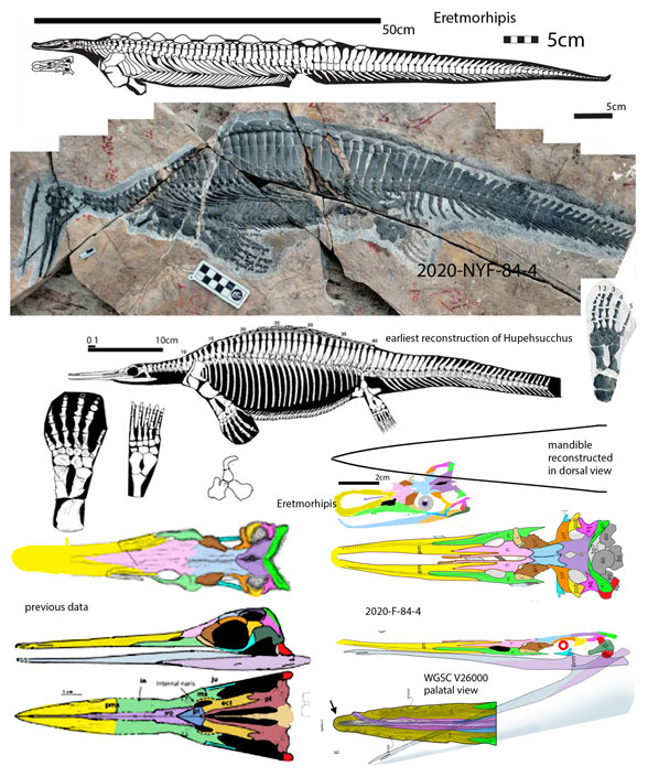
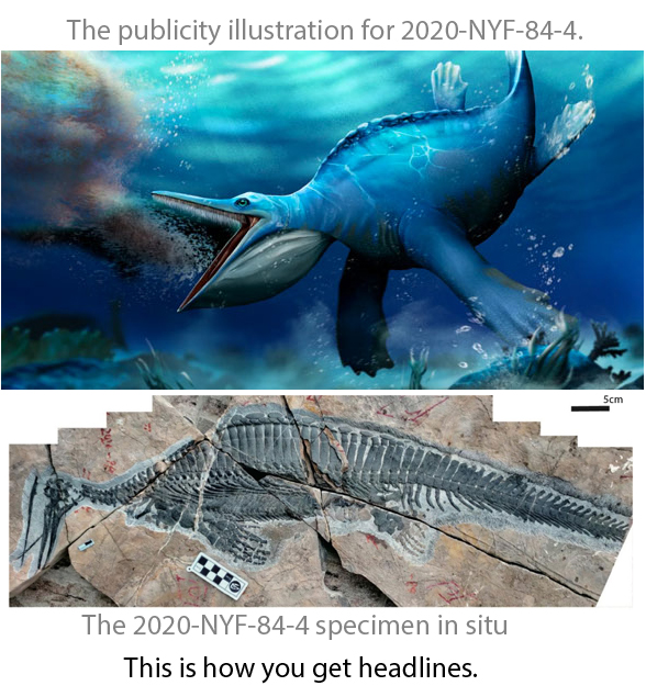
Figure 1. Huphesuchus, the 2020-F-84-4 specimen and Eretmorohipis compared. Note the medial fenestra and laterally oriented dorsal ribs in Eretmorhipis and the 2020-F-84-4 specimen. Look at the rostrum of the 2020-F-84-4 speicmen. Is it more like a kitchen strainer? Or more like a tweezers?
Unfortunately, when the new 2020-F-84-4 specimen was added to the LRT, it did not nest with Hupehsuchus (Fig 1), but closer to Eretmorhips(Fig 1).
Unfortunately the unfused upper jaw and divided premaxilla of 2020-F-84-4 is not shared with related taxa. This condition appears to be due to taphonomic damage at the tip. Undamaged specimens (Fig 1) retain that tip that bridges both sides of the premaxilla.
Unfortunately the ‘specialized intermediate space’ is shared with Eretmorhipis (Fig 1), not Hupehsuchus. Another related taxon, Eohupehsuchus, has an even narrower rostrum lacking any dorsomedial opening.
From the Fang et al abstract: “Hupehsuchus had enlarged its buccal cavity to enable efficient filter feeding and probably used soft tissues like baleen to expel the water from the oral cavity.”
The buccal cavity (= mouth) or the 2020-F-84-4 specimen is actually relatively smaller compared to its body than the holotype of Hupehsuchus (Fig 1). However, the mouth is indeed larger on the publicity illustration (Fig 2).
The buccal cavity of 2020-F-84-4 can be described as small, flat and pointed. Is there room for baleen-like filtering structures in the extremely shallow rostrum of the 2020-F-84-4 specimen? Maybe not. Are those needle-thin mandibles capable of bowing out like mysticete and pelican jaws? Maybe not. Usually if a taxon is gulping a lot of water, those jaws are really big. The jaws on this and related hupehsuchids are more like flat to pointed tweezers. They are particularly gracile on 2020-F-84-4 (Fig 1).
Figure 2. Two images of the same specimen. One for science. One for publicity. If you see any differences or exaggerations, so does everyone else. If you want your specimen to have a larger mouth rimmed with baleen and more robust limbs, just let the illustrator know. Is this good science? You decide.
The authors mention studying one platypus, which has a similar flat toothless, baleen-less buccal cavity, a similar overall morphology, a similar size and thus more likely a similar lifestyle. No further mention was made of that platypus in the text and its similarity to hupehsuchids in general. We looked at this topic a few years ago here.
From the Fang et al abstract: “Coordinated with the rigid trunk and pachyostotic ribs suggests low speeds of aquatic locomotion, Hupehsuchus probably employed continuous ram filter feeding as in extant bowhead and right whales.
Maybe not. Their figure 2 compares Hupehsuchus to a rorqual, a Minke whale, not a right or bowhead whale. If you say ‘right whale’ you should show ‘right whale’. If you show a Minke whale, you should say a MInke whale. And show them to scale
The Early Triassic palaeoenvironment of a restrictive lagoon with low productivity drove Hupehsuchus to feed on zooplankton, which facilitated ecosystem recovery in the Nanzhang-Yuan’an Fauna at the beginning of the Mesozoic.”
When a changing niche drives a taxon to a particular modification, you’re going to have to show some ancestors and some phylogenetic relatives. As mentioned earlier, Early Permian pre-mesosaurs were ancestral to hupehsuchids + ichthyosaurs in the LRT. So there’s a starting point worth examining, but overlooked by the authors.
Take a look at Wumengosaurus (Fig 3) for a hupehsuchid relative.
Another reason to run an analysis: Make sure your new taxon is indeed the genus and species you call it in your headline. This time a mistake was made.
Figure 3. From 2018. Basal Ichthyosauria, including Wumengosaurus, Eohupehsuchus, Hupehsuchus and Thesaurus, the taxon closest to ichthyosaurs.
What does a platypus eat? “They scoop up insects and larvae, shellfish, and worms in their bill along with bits of gravel and mud from the bottom. All this material is stored in cheek pouches and, at the surface, mashed for consumption. Platypuses do not have teeth, so the bits of gravel help them to “chew” their meal.”
Sounds like what a hupesuchid might do, even without baleen.
References Fang et al (8 co-authors including testbook author MJ Benton) 2023. First filter feeding in the Early Triassic: cranial morphological convergence between Hupehsuchus and baleen whales. BMC Ecology and Evolution. https://doi.org/10.1186/s12862-023-02143-9
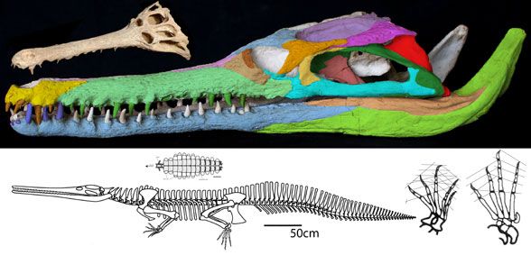
This is the dinosaur that started the dinosaur renaissance. Deinonychus antirrhopus(Ostrom 1969, Early Cretaceous, Figs 1–4) had the first ‘killer’ claw, a tendon stiffened tail and an Archaeopteryx-like manus (hand). These traits indicated a highly active, likely warm-blooded predator, more like an osprey (Pandion), than a Komodo dragon (Varanus).
Figure 1. The Harvard specimen of Deinonychus. This is not a complete specimen. Some preserved parts are detailed below.
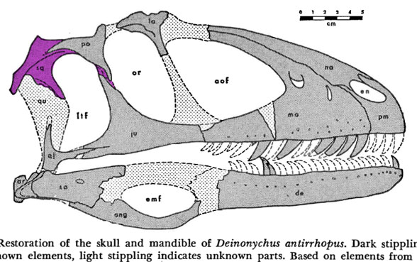
Figure 2. Deinonychus skull. Colors added here. Note the long jugal-squamosal (cyan-magenta) contact.
Figure 2b. Figure from Ostrom 1970. Color added here to show the extent of the squamosal. Any differences between the original drawings and the sculptured skull are in the purview of the museum sculptors, who appear to have had access to the original materials.
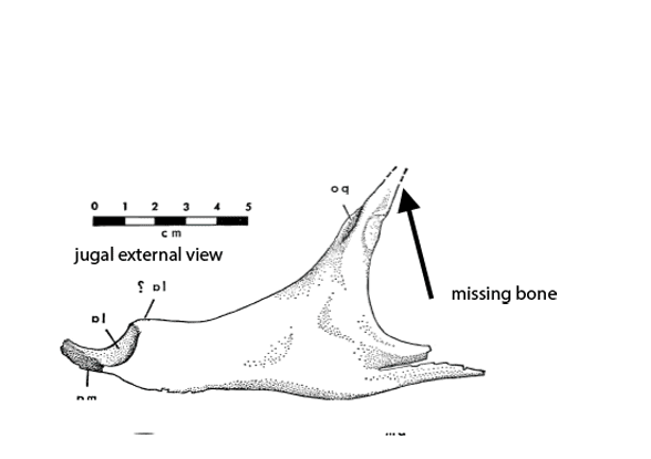
Figure 2c. A preparator’s dilemma. Did the jugal contact the squamosal or not? The preparator made a choice to show contact (Fig 2). Ostrom’s 1970 drawing of the individual bones (assembled here) indicates vanishing bone at the key area. Here two possibilities for contact are shown. Of course, bone could have been absent here – but the model maker chose contact from data known to them. As readers know, corrections are part of the scientific process. This is correction #212,444 or thereabouts.
Distinct from tested relatives the orbit of Deinonychus (Fig 2) is deeper than wide and not wider than the post-orbital length of the skull. Note the long jugal-squamosal (cyan-magenta) contact is also distinct.
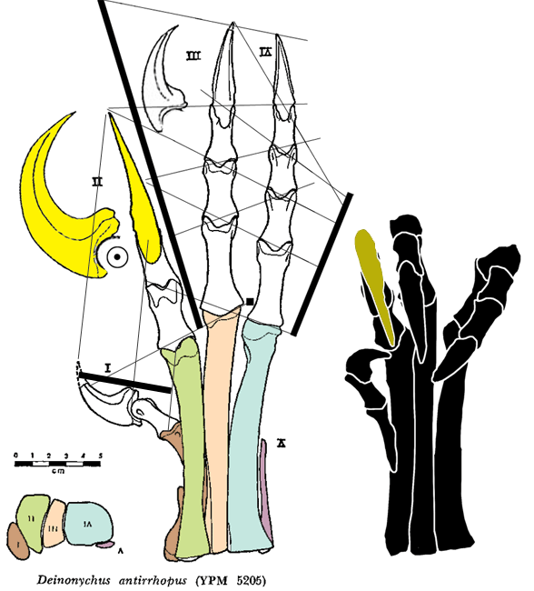
PILs (parallel interphalangeal lines) can be drawn through most, but not all of the phalangeal joints of the hand (Fig 3) and foot (Fig 4).
Figure 4. Deinonychus pes, extended and flexed in ventral view. Parallel Interphalangeal Lines (PILs) added here.
In the LRT Deinonychus is derived from Late Jurassic Ornitholestes and Late Jurassic Sciurumimus, neither of which had a ‘killer’ claw nor a chevron-tendon-stiffened tail.
References Ostrom JH 1970. Stratigraphy and paleontology of the Cloverly Formation (Lower Cretaceous) of the Bighorn Basin area, Wyoming and Montana”. Bulletin of the Peabody Museum of Natural History. 35: 1–234.