Beishanichthys and Amia: related or not?

The extant bowfin (Amia) nests as a semi-basal ray-fin fish
in the large reptile tree (LRT, 2227 taxa), a descendant of Middle Triassic Fukangichthys (Fig 1) and Early Triassic Beishanichthys (Fig 1). That answers the headline question.

By contrast, Xu and Gao 2011 described Beishanichthys
as a ‘scanilepiform’. “Contrary to previous thought that scanilepiforms are closely related to
the Amiidae, the phylogenetic results of this study recognize the Scanilepiformes as stem-group neopterygians. Relationships of the Scanilepiformes and Australosomus with other neopterygians remain unresolved.”

According to Xu and Gao, “The Scanilepiformes Sytchevskaya, 1999 are an extinct group of ‘palaeoniscoid’ fishes with a geological range confined to the Triassic.”

According to the LRT, Beishanichthys is not a palaeoniscoid (= close relatives of Palaeoniscum). Taxon exclusion due to academic tradition is the problem here. The LRT minimizes taxon exclusion by including so many more taxa.

Scanilepis (Fig 3), a recent addition to the LRT prompted by these taxa, nests with Beishanichthys (Fig 1), close to Amia (Fig 1), not related to Palaeoniscum.

Figure 1. Bowfin (Amia) ancestors in the LRT to scale.
Figure 1. Bowfin (Amia) ancestors in the LRT to scale. Here the resemblance between Beishanichthys and Amia is not just superficlal, it is due to a close interrelationship.

Meanhwhile, in their description of Fukangichthys,
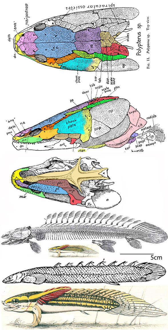
(Fig 1) Giles et al 2017 reported, “We show that scanilepiforms, a widely distributed Triassic (ca. 251-200 Mya) radiation, are stem polypterids. Polypterids (bichirs and ropefish) represent the earliest-diverging lineage of living actinopterygians.”

By contrast, in the LRT Polypterus is a basal lungfish, not related to Fukangichthys or early diverging actinopterygians (= ray-fin fish). Polypterus is a lobe-fin. Moreover, Scanilepiformes (1999) appears to be a junior synonym for Amiformes (1929).

Unfortunately
Prior workers were working under the academic tradition that Amia was a member of the invalid clade, Chondrostei, which traditionally includes bichirs, sturgeons and spoonbills. In the LRT none of these taxa are related to one another. In the LRT Amia, Beishanichthys and Fukangichthys (Fig 1) are closely related basal ray-fin fish (Fig 2).

Figure 2. Extant basal ray-fin fish in the LRT include Engraulis, Malacosteus and Dactylopterus. All three share the trait of a large lateral orbit close to the short rostral tip.

Amia calva
(Linneaus 1766; up to 70cm in length) is the extant bowfin, a basal ray-fin fish able to breathe both water and air. As in related Beishanichthys, a single elongate undulating fin is present. Hatchlings look like tadpoles. Fossil relatives of Amia have a worldwide distribution in fresh and salt waters.

Figure 3. Scanilepis from Lehman 1979. Colors added here.
Figure 3. Scanilepis from Lehman 1979. Note the differences between the Lehman diagram and the fossil. Colors added here.

Scanilepis dubius
(Lehman 1979, Late Traissic, 1.6m long) is a much larger relative of Beishanichthy and the living bowfin, Amia. Note the long dorsal fin and short rostrum. These related taxa indicate the large postorbitals of Amia are the result of fusion between the postorbitals (amber) and jugals (cyan).

This appears to be a novel hypothesis of interrelationships.
If not please provide a citation so I can promote it here.

References
Giles S, Xu G-H, Near TJ and Friedman M 2017. Early members of ‘living fossil’ lineage imply later origin of modern ray-finned fishes. Nature. 549 (7671): 265–268.
Lehman JP 1979. Le genre Scanilepis Aldinger du Rhétien de la Scanie. Bulletin of the Geological Institutio n of the University of Uppsala, N.S 8:113-125.
Linneaus C von 1766. Systema naturæ per regna tria naturæ, secundum classes, ordines, genera, species, cum characteribus, differentiis, synonymis, locis. Tomus I. Editio duodecima, reformata. pp. 1–532. Holmiæ. (Salvius) .
Su T 1978. Memoirs Inst. Vert. Paleont. Paleoanthrop. Peking No. 13.
Xu G-H and Gao K-Q 2011. A new scanilepiform from the Lower Triassic of northern Gansu Province, China, and phylogenetic relationships of non-teleostean Actinopterygii PDF. Zoological Journal of the Linnean Society. 161 (3): 595–612.
Xu G-H, Gao K-Q and Finarell JA 2014. A revision of the Middle Triassic scanilepiform fish Fukangichthys longidorsalis from Xinjiang, China, with comments on the phylogeny of the Actinopteri. Journal of Vertebrate Paleontology 34(4):747–759.

wiki/Amia
wiki/Fukangichthys
wiki/Beishanichthys
wiki/Scanilepis

Scottish Middle Jurassic Borealestes enters the LRT next to a Late Triassic basal cynodont, Prozostrodon. Those mammal-like teeth were by convergence.

Pancirolli et al 2016 reported,
“The Middle Jurassic docodont Borealestes serendipitus was the first Mesozoic mammal found in Scotland over 40 years ago. Its affinities and morphology have remained poorly understood. We present an updated description and diagnosis of the genus Borealestes, based on high-resolution micro-computed tomography (micro-CT) and synchrotron scans. Our phylogenetic analysis supports a clade formed by Borealestes, Haldanodon, Docofossor, and Docodon. Ontogenetic variation in the mandibular morphology of Borealestes is similar to that seen in Docodon and Haldanodon, with the delayed emergence of the ultimate lower molar, the shift of the last molar to the front of the coronoid process, and a posterior shift of the Meckel’s sulcus in successively older individuals. This supports a distinctive growth pattern in the clade including Borealestes and Docodon, one that may be present in Docodonta as a whole.”

Borealestes and Haldanodon are not mammals according to the large reptile tree (LRT, 2226 taxa). The were late-surviving [Jurassic] probainognathid cynodonts that independently developed mammal-like teeth (Fig 1) and lots of them in tiny Borealestes.

Figure 1. Borealestes restored and mandible from Panciroli et al 2016. Tooth colors added here.  The authors indicated a double-rooted molar, here re-identified as a first premolar.
Figure 1. Borealestes restored and mandible from Panciroli et al 2016. Tooth colors added here. The authors indicated a double-rooted molar, here re-identified as a two-rooted worn down first premolar.

According to Wikipedia,
“Docodonta is an order of extinct mammaliaforms that lived during the Mesozoic, from the Middle Jurassic to Early Cretaceous. They are distinguished from other early mammaliaforms by their relatively complex molar teeth.”

The putative clade, ‘Mammaliaformes is a junior synonym for ‘Mammalia’ since it relies on the basal marsupial, Morganucodon, as an outgroup.

Middle Triassic Prozostrodon (Fig 2) was not tested, nor mentioned in the text.

The most interesting thing about this taxon
is the parallel evolution of mammal-like teeth into the Middle Jurassic. There were more teeth than any mammal. Here the double-rooted canine is reinterpreted as the first of six premolars. The single-rooted canine was probably no larger than the incisors, based on its diameter.

Figure 2. The cappa-ufsm-021 specimen of early Late Triassic Prozostrodon compared to size and to scale with Borealestes.
Figure 2. The cappa-ufsm specimen of early Late Triassic Prozostrodon compared to size and to scale with two specimens of the smaller Middle Jurassic Borealestes.

Co-author, Dr. Elsa Panciroli [Oxford Museum of Natural History]
is also the author of ‘Beasts Before Us, The untold story of mammal evolution‘. She discussed the book’s contents on YouTube as part of the Royal Tyrrell Museum of Paleontology series.

Actually that ‘untold story’ was told thirty years ago
in ‘From the Beginning, the Story of Human Evolution‘ (Wm Morrow 1991), and is further expanded with more taxa in the LRT, 2226 taxa), and probably in every college textbook.

Panciroli defines mammals
with lactation and three inner ear bones, rather than the preferred last common ancestor method (e.g. Megazostrodon, Ukhaatherium, their last common ancestor and all of its descendants).

Panciroli gives herself credit for moving that origin of mammals
to 250mya (in the video) to 300 mya (in the caption). This is not news.

According to the author,
the first group to split off from ‘the amorphous taxa’ were the amphibians (= the anamniotes according to the caption). She says, “Amniotes laid eggs with a tough, leathery shell.” Not necessarily. The shell is another matter. The amnion is the key membrane.

Unfortunately
the author does not realize the actual term is “Reptile”. Amniote is a junior synonym.

More textbook mythology follows.
The author reports the first split was between the synapsids and diapsids. This is wrong on at least two levels. All basal reptiles had no skull openings. Diapsid architecture evolved twice, once from synapsid ancestors.

The author thinks the story of pelycosaurs
“is missed out of our telling, not just of mammal history, but really history of life on Earth.” She focuses on Dimetrodon.

This is wrong. Dimetrodon is not part of the mammal story. We knew this thirty years ago. She claims Dimetrodon is not a reptile. This is wrong. All animals with an amniotic membrane are reptiles. Where is the concept of monophyly? Dr. Panciroli is stuck in the 1960s.

Next Panciroli describes therapsids as the most diverse lineage
in the Permian, wasting time with dicynodonts. This is supposed to be the untold story of mammal evolution. She mentions scale-less skin preservation in Estemmenosuchus, not realizing that all early amniotes and many Permian taxa lacked scales plesiomorphically. Scales appeared in the Triassic, spurred by the rise of biting insects.

After 34 minutes
Panciroli discussed her first mammal ancestor, Procynosuchus.

At 35 minutes Dr. Panciroli describes Thrinaxodon,
with a complex and branched maxillary canal, indicative of whiskers. Again, this information is at least thirty years old. Panciroli attributes milk production to Thrinaxodon, because it loses the pineal opening. This is wrong. Single replacement teeth indicate milk production. So does the last common ancestor method, which attributes milk production to nothing before monotremes.

Panciroli reported mammals begin to evolve smaller sizes during the Mesozoic.
This is only occasionally true (e.g. Hadrocodium), but most early mammals tested in the LRT were mouse-to squirrel-sized. I only hope the book is better than the teaser talk.

Correctly Panciroli describes
the reduction of posterior jaw bones to ear bones. This is not news.

At 41:00 Panciroli acknowledges in general terms
a diverse assemblage of Mesozoic mammals, none larger than a badger this time.

At 43:00 the author describes Early Cretaceous Repenomamus,
not realizing it was a late-survivng, pre-mammal cynodont, not a mammal.

At 45:00 Panciroli discusses the asteroid
at the K-Pg boundary. So out of 50 minutes, only ten were given to the ‘untold’ Mesozoic mammal story. No time was given to the Cenozoic taxa. For the remaining 32 minutes viewer questions were answered.

Suggestion to Dr. Panciroli:
Build your own LRT so you can understand the evolution of mammals and their untold story. It’s not good to cherry-pick a few well-worn tales published years ago and tell readers, your book is the ‘untold story’.

References
Pancirolli E, Benson R and Luo Z-X 2016. The mandible and dentition of Borealestes serendipitus (Docodonta) from the Middle Jurassic of Skye, Scotland. Journal of Vertebrate Paleontology 39(3):Article: e1621884
Waldman M and Savage RJG 1972. The first Jurassic mammal from Scotland”. Journal of the Geological Society. 128 (2): 119–125.

wiki/Borealestes
wiki/Docodonta
wiki/Mammaliaformes
youtube.com/watch?v=ukJfEt0Jh6E

Why multituberculates went extinct in the Late Eocene

The longest lived minor clade of placental mammals, Multituberculata
(Middle Jurassic to Late Eocene), mysteriously went extinct when similar placentals did not. Perhaps this was due to the rise of carnivorous raptorial birds (e.g. hawks, eagles, owls).

This is a novel hypothesis based on timing and hearing.

Figure 1. Animation of the mandible of the multituberculate Kryptobaatar showing the sliding of the jaw joint producing separate biting and grinding actions, just like rodents, their closest relatives in the LRT.

As long-time readers might recall
multituberculates evolved from rodents and plesiadapiformes in the large reptile tree (LRT, 2225 taxa), the only cladogram to test and nest these three clades together.

Due to the sliding jaw joint of multis
(Fig 1) their three tiny ear bones failed to develop into the exquisitely sensitive tiny middle ear bones of their relatives. Chalk that up to a reversal via neotony. Their post-dentary bones stopped developing at the embryo stage and remained large elements attached to the dentary as an adult, like those of pre-mammals. Many arboreal therians recapitulate phylogeny during ontogeny like this.

That’s the only reason why many paleontologists consider multis to be at least as primitive as monotremes, if not more primitive. That’s why they don’t test multis with rodents and plesiadapiformes. The LRT is not limited by such assumptions.

Figure 3. Comparing multituberculate origins: Cziki-Sava et al. vs. LRT.
Figure 2. Comparing multituberculate origins: Cziki-Sava et al. vs. LRT.

Poor hearing was fine so long as
multis had the trees largely to themselves. However, by the Eocene a new clade of predators capable of snatching arboreal mammals out of the branches came along.

Figure 6. The aye-aye, Daubentonia in vivo. This is the closest living relative of multituberculates and is itself a plesiadapiform member of Glires, close to rodents, not primates.
Figure 3. The aye-aye, Daubentonia in vivo. This is the closest living relative of multituberculates and is itself a plesiadapiform member of Glires, close to rodents, not primates.

Hawks didn’t care whether their prey was
a rodent, a plesiadapiform, a primate or a multituberculate. Hawks attacked them all. Unfortunately multis were then at a disadvantage, due to their less acute hearing. As a result they were relatively ‘easy pickins’ and their numbers dwindled to extinction. Only rodents, with their excellent hearing, managed to survive worldwide. Small primates (including lemurs and adapids) disappeared except in heavily jungled areas near the equator. Plesidapiformes survived as a single nocturnal jungle taxon, Daubetonia, the aye-aye, on Madagascar, along with native (not rafting) lemurs. Owls also hunt on Madagascar, so it wasn’t the mere presence of nocturnal hunting birds that doomed arboreal mammals. It was the newly evolved hunting birds > plus < the less acute hearing of multis that hastened their undoing.

Or is there a better explanation? Let me know if you have one.

Van Valen and Sloan 1966 wrote:
“The multituberculate mammals declined in numerical abundance from the late Cretaceous to their extinction in the late Eocene. Their maximal diversity, however, occurred in the late part of the middle Paleocene, suggesting an increase in specialization in the face of competition from placental mammals, which diversified very rapidly in the Paleocene. Various lines of evidence lead to the conclusion that first condylarths, then primates, and finally rodents contributed to the gradual extinction of multituberculates, at least in part by evolution in the same region of competing resource requirements.”

Maas, Krause and Strat 2016 echoed Krause 1986 when they wrote:
“Analyses of body size, diet, activity patterns, and locomotion support the hypothesis that rodents and non-paromomyid plesiadapoids may have competed for the same resources.”

Like other workers, these authors do not mention poor hearing and hawks.

References
Krause, DW 1986. Competitive exclusion and taxonomic displacement in the fossil record: the case of rodents and multituberculates in North America. Pp. 95–117. In Flanagan, K. M., and Lillegraven, J. A. (eds.), Vertebrates, Phylogeny and Philosophy. Contributions to Geology Special Paper 3. The University of Wyoming; Laramie, Wyoming.
Maas M, Krause DW and Strait SG 2016. The decline and extinction of Plesiadapiformes (Mammalia: ?Primates) in North America: displacement or replacement? Paleobiology 14(4):
Van Valen L and Sloan RE 1966. The extinction of the multituberculates. Systematic Zoology 15:261–278.

New ‘whale’ ancestor paper

Burin et al 2023 looked at
mysticete and odontocete body length, then added fossil taxa to document trends in ‘whale’ size.

Figure 1. Odontoceti (toothed whale) origin and evolution. Here Anagale, Andrewsarchus, Sinonyx, Hemicentetes, Tenrec Indohyus and Leptictidium precede Pakicetus. Maiacetus and Orcinus are aquatic odontocetes.
Figure 1. Odontoceti (toothed whale) origin and evolution. Here Anagale, Hemicentetes, Tenrec Indohyus and Leptictidium precede Pakicetus. Maiacetus and Orcinus are aquatic odontocetes. This is an older illustration. Adding taxa moved Andrewsarchus and Sinonyx away from this clade.

Unfortunately,
Burin’s team still holds to the invalid hypothesis that mysticetes and odontocetes are related to one another. This is a myth that has to go away. Several years ago the large reptile tree (LRT, 2224 taxa) falsified the hypothesis of monophyly in the ‘Cetacea’ by simply adding taxa.

Figure 1. Taxa in the lineage of right whales include Desmostylus, Caperea and Eubalaena. The tiny bit of jugal posterior to the orbit (in cyan) is found in all baleen whales tested so far. The frontals over the eyes are just roofing the eyeballs in Desmostylus, much wider in Caperea and much, much longer in Eubalaena.
Figure 2. Taxa in the lineage of right whales include Desmostylus, Caperea and Eubalaena. The tiny bit of jugal posterior to the orbit (in cyan) is found in all baleen whales tested so far. The frontals over the eyes are just roofing the eyeballs in Desmostylus, much wider in Caperea and much, much longer in Eubalaena.

According to phys.org/news:
“To get a more detailed look at cetacean evolution [ there is no such thing as a ‘cetacean’], a team of scientists, including Dr. Travis Park and Dr. Gustavo Burin, both Leverhulme postdocs at the Natural History Museum, gathered body length measurements of 345 different species, including 89 living species and 256 fossil lineages, in the largest dataset of its kind. Their article, “The Dynamic adaptive landscape of cetacean body size” is published in Current Biology.”

“By comparing body lengths from both living and fossil species—an approach rarely taken—the research revealed that the evolutionary trends in cetacean size remained much the same for over 20 million years after they entered the ocean.”

Mysticetes and odontocetes entered the ocean separately.

“The first cetaceans were goat-sized animals which lived on the edge of lakes and rivers, spending time both in and out of water. Over time, their descendants became increasingly adapted to life in water, before leaving land behind altogether.”

Since these authors have no idea that mysticetes evolved by convergence with mysticetes, this paper becomes a waste time for the writers, editors, referees and readers – unless it is split into two works. Readers, make sure your phylogeny is valid.

References
Gustavo Burin et al 2023. The dynamic adaptive landscape of cetacean body size, Current Biology (2023). DOI: 10.1016/j.cub.2023.03.014

https://phys.org/news/2023-03-fossils-whale-ancestors-reveals-path.html

Origin of the squamosal in flat-head, pre-tetrapod fish

The earliest (= most primitive) bony fish
had a jugal (cyan in figure 1), but no squamosal (magenta in figure 3) tetrapod homolog. The squamosal first appeared in flat-head, pre-tetrapod fish like Grossius and Tinirau (Fig 3) between splitting jugal elements that were a large single element in Eusthenopteron (Fig 2).

Figure 1. Tarrasius is a small Early Carboniferous fish basal to extant moray eels. Note the presence of a jugal (cyan), but a lack of a squamosal (magenta cheek bone) as seen in figure 2.

The recognition of the splitting of the jugal
with the appearance of the squamosal is one of those vexing things that made this phylogenetic study of fish extend as long as it has. This was vexing because traditional labels changed.

Figure 3. Eusthenopteron was originally labeled with a squamosal (cyan here), but here that bone is a large jugal and a squamosal is not present. The two tan bones are lacrimals. The posterior lacrimal is its way out after original attachment to the maxilla. The tetrapod lacrimal (anterior tan element here, splits off from the circumorbital ring, which were originally postorbital elements (amber).
Figure 2. Eusthenopteron was originally labeled with a squamosal (cyan). Here that bone is a large jugal and a squamosal is not present. The two tan bones are here labeled lacrimals. The posterior lacrimal is on its way out after original detachment from the maxilla (see figure 1). The tetrapod lacrimal (anterior tan element here) is from the circumorbital ring, originally all postorbital elements (amber). Compare to figure 1. The anterior lacrimal migrates closer to the posterior naris, a pre-choana that migrates to the jaw margin, then moves on to the palate, disappearing from the exterior in pre-tetrapods. Compare this skull to Selenodus in figure 3. The lacrimal precedes the premaxilla and maxilla, forming a base for both to develop on in certain placoderms, sharks and acanthodians.

Ray-fin fish never developed a squamosal
and neither did many lobe fins. Starting with Late Devonian Grossius, a lobe-fin with a wider-than-tall skull (Figs 3, 4), a squamosal first appears and is largely retained in tetrapods.

Figure 1. Left: Grossius demonstrates the origin of the squamosal (magenta), splitting the twin jugals. The upper jugal eventually disappears. Right: Selenodus shows the plesiomorphic (pre-squamosal) condition.
Figure 3. Left: Grossius demonstrates the origin of the squamosal (magenta), splitting the twin jugals. The upper jugal eventually disappears. Right: Selenodus shows the plesiomorphic (pre-squamosal) condition. Schultze 1973 thought both of these taxa were related to Onychodus. In the LRT only Selenodus is related to Onychodus.

Grossius aragonensis
(Schultze 1973; Late Devonian) has a straight rostrum and a wider-than-tall skull. Note the splitting of the jugal as the squamosal (magenta) moves the two jugal elements apart.

Figure 2. Grossius compared to Middle Devonian Tinirau, a flatter pre-tetrapod, pre-Panderichths taxon.
Figure 3. Grossius compared to Middle Devonian Tinirau, a flatter pre-tetrapod, pre-Panderichths taxon. The splitting of the jugal is just one of many things you learn > after < studying fish skulls for 5 months. Sorry it took so long. I started as a freshman on this topic and the labels were misleading.

Tinirau clacke
(Swartz B 2012; Middle Devonian; UCMP 118605) nests between Cabonnicthys and Panderichthys in the LRT. The skull is twice as wide as tall, the skull bones are more tetrapod-like, dorsal ribs make a first tentative appearance. The tail was likely straight, as in sister taxa.

This hypothesis re-labels fish skull bones with tetrapod homologs
to facilitate tracing their development across all clades and taxa (Figs 1–3) in the LRT. I wish this practice was more widespread. Traditional academics also label many fish skull bones this way, but not to this extent. They have their own nomenclature.

For example: the traditional fish preorbital is a tetrapod homolog postorbital (the entire circumorbital ring, other than the postfrontal). In one clade a portion becomes the tetrapod lacrimal. Other examples are seen in figure 2.

The preopercular and opercular are not found in tetrapods, nor are they transformed during evolution into new bones. They just disappear.

This appears to be a novel hypothesis of fish skull bone homologs.
If not, please provide a citation so I can promote it here.

References
Schultze H-P 1973. Crossopterygier mit heterozerker Schwanzflosse aus dem Oberdevon Kanadas, nebst einer Beschreibung von Onychodontida-Resten aus dem Mitteldevon Spaniens und aus dem Karbon der USA. Palaeontographica, Abteilung A, 143:188–208.
Swartz B 2012. A marine stem-tetrapod from the Devonian of Western North America. PLoS ONE. 7 (3): e33683. doi:10.1371/journal.pone.0033683

wiki/Tinirau
wiki/Grossius

A Cretaceous gonorynchid, Sapperichthys, also enters the LRT with sturgeons

Amaral et al 2013 described
Sapperichthys (Fig 1) as a new gonorynchid from the Cenomian (earliest Late Cretaceous) of Chiapas, Mexico. The authors did not consider sturgeons in their phylogenetic analysis, nor did they mention sturgeons in their text.

Figure 1. Sapperichthys from Amaral et al 2013. Colors and reconstruction added here.
Figure 1. Sapperichthys from Amaral et al 2013. Rostrum and tail lost during excavation. Colors and reconstruction added here. In situ fossil shown about actual size on 72dpi monitors. Note the diamond-shaped dorsal armor in the fossil, as in Acipenser. See figure 2.

Here
in the large reptile tree (LRT, 2224 taxa) Sapperichthys nests with sturgeons, between adult sturgeons and the larval Acipenser sturgeons (Fig 1) + Gonorynchus + Cromeria.

Figure 2. Acipenser brevirostrum, 1m typical length. Records up to 1.47m.

This appears to be a novel hypothesis of interrelationships
overlooked due to taxon exclusion. If not please provide a citation so I can promote it here.

References
Amaral CRL, Alvarado-Ortega J and Briot PM 2013. Sapperichthys gen. nov., a new gonorynchid from the Cenomanian of Chiapas, Mexico. In: Mesozoic Fishes 5 – Global Diversity and Evolution, Arratia, Schultze and Wilson (eds.): 305–323. by Verlag Dr. Friedrich Pfeil, München, Germany – ISBN 978-3-89937-159-8

wiki/Sapperichthys – not yet posted

The wide-set eyes of the marsupial sabertooth, Thylacosmilus

Gaillard, MacPhee and Forasiepi 2023 reported,
“Adaptations for good stereopsis have evolved in living predaceous mammals, and it is reasonable to infer that fossil representatives would follow the same pattern. This applies to the sparassodonts, an extinct group of South American hypercarnivores related to marsupials, with one exception. In the sabertooth Thylacosmilus atrox, (Fig 1) the bony orbits were notably divergent, like those of a cow or a horse, and thus radically differing from conditions in any other known mammalian predator.”

Not true. ‘Divergent orbits’ (Fig 1) is a trait common to clade members (e.g. Fig 4), as one should expect in any clade. Unfortunately phylogenetic analysis was not part of this study and several marsupial sabertooth clade members were ignored.

Readers, whenever you see the term, “radically differing”, assume the authors have not done their homework (or might be seeking headlines). Evolution never produces anything radically different. It always works in baby steps (= microevolution). Relatives share traits.

Sprassodonta is polyphyletic according to the large reptile tree (LRT, 2224 taxa) with some traditional members nesting in Placentalia, others in Marsupialia.

Figure 2. Thylacosmilus skull. Colors added here. Compare to Barbourofelis in figure 1.Figure 2. Thylacosmilus skull. Colors added here. Compare to Barbourofelis in figure 1.
Figure 1. Thylacosmilus skull. Colors added here. Note the wide orientation of the orbital axes, the subject of the authors’ paper. Note the deep canine roots, further splitting the orbits from each other. This trait is also found in Early Cretaceous Vincelestes (see figure 2). Compare this skull to that of Patagosmilus in figure 2 and Barbourofelis in figure 3.

The authors’ report,
“The factors that prompted orbital reorganization in Thylacosmilus are unknown…” that may be because only one other ancestral taxon was listed in the text (house-cat sized Patagosmilus, Fig 2), but not illustrated. The authors could have answered this question by testing ancestral and related taxa. They did not do so. In the LRT this sabertooth marsupial clade goes back in time to rat-sized Early Cretaceous Vincelestes (Fig 2). Even deeper in this lineage are extant Dasycercus and Dasyuroides (Fig 6), also not mentioned in the text. The latter is known to, “kill with strong bites to the head” of its victims. Phylogenetic bracketing indicates that extant habit could help us understand the habits of the extinct related Thylacosmilus (Fig 1).

The authors > also < report,
“The forcing function behind these morphological tradeoffs was the extraordinary growth of its rootless canines, which affected skull shape in Thylacosmilus in numerous ways, including relative orbital displacement.”

So, “The factors that prompted orbital reorganization in Thylacosmilus” are known, according to the authors. This is easy to see (Fig 1). The question the authorsleave unanswered (due to taxon exclusion) is: how far back does this trait extend?

Figure 4. Patagosmilus and Vincelestes at full scale on a 72dpi monitor. These are derived members of the sabertooth marsupial clade.
Figure 2. Eocene Patagosmilus and Early Cretaceous Vincelestes at full scale on a 72dpi monitor. In the LRT these are members of the sabertooth marsupial clade that includes Thylacosmilus.

Gaillard, MacPhee and Forasiepi reported,
“We show that the orbits of Thylacosmilus were frontated and verticalized in a way that favored some degree of stereopsis and compensated for limited convergence in orbital orientation.

Figure 5. Skull of Barbourofelis, a traditional nimvravid placental cat, nests in the LRT with Thylacosmilus after trait analysis.
Figure 3. Skull of Barbourofelis, a traditional nimvravid placental cat, nests in the LRT with marsupial Thylacosmilus after trait analysis. Note the similar deep roots that rotate the orbits further laterally.

This can’t be the first time
this ‘limited convergence in orbital orientation’ has been noted. Or is it? Have these three authors finally unveiled the obvious? Or are they making a big deal out of something everyone is already aware of?

Figure x. Smilodon skull, skeleton, manus and pes with PILs added.
Figure 4. Smilodon skull, skeleton, manus and pes with PILs added. Compare to Stylinodon in figure y.

Barbourofelis
(Fig 3) is traditional nimravid (= placental cat), but in the LRT nests with the marsupial, Thylacosmilus (Fig 1), sharing a similar skull architecture not found in placental cats (including Nimravus Fig 5) or Smilodon, Fig 4). To force Barbourofelis to move in the LRT requires 28 additional steps to Smilodon, and 34 additional steps to Nimravus.

Figure 4. Osbornudon to scale with Nimravus.
Figure 5. The nimravid cats, Osbornudon to scale with Nimravus.

Phylogenetic analysis answers so many questions.
Build your own LRT so you can use this powerful tool to answer phylogenetic questions like those Gaillard, MacPhee and Forasiepi both posed and overlooked.

Figure 8. Extant Dasyuroides is a carnivorous marsupial in the lineage of Thylacosmilus.
Figure 6. Extant Dasyuroides is a carnivorous marsupial in the lineage of Thylacosmilus.

References
Gaillard C, MacPhee, RDE and Forasiepi AM 2023. Seeing through the eyes of the sabertooth Thylacosmilus atrox (Metatheria, Sparassodonta). Nature communications biology https://doi.org/10.1038/s42003-023-04624-5

wiki/Sparassodonta

Theropod lips? Don’t forget Sinornithosaurus.

“Animals like T-Rex, theropod dinosaurs, most likely had some sort of lips, like a soft tissue covering on their mouth to cover their teeth,” said one of the authors of the study, Thomas Cullen, an assistant professor of paleobiology at Auburn University.

As everyone knows, living theropods (birds) and living archosaurs (crocs) lack lips. On the other hand, lizards (including snakes) have lips, as do most tetrapods.

That sets the stage for this argument. Cullen et al 2023 decided to forsake phylogenetic bracketing and place their bets on convergence with lizards and their similar skull foramina.

Cullen et al also chose to illustrate their hypothesis
by using one Tyrannosaurus with relatively small teeth (Fig 1, FMNH PR 2081), rather than really test their hypothesis with several theropods with much larger lateral teeth (Figs 2, 4).

If Cullen et al were trying to set a rule, that rule should apply to one and all.
More toothy specimens (Figs 2, 4) were not illustrated in Cullen et al.

Figure 1. T- rex illustrations from Cullen et al 2023.
Figure 1. T- rex illustrations from Cullen et al 2023. Note the authors are indicating at least half of the upper tooth coverage comes from the lower lip. Their mismatch of the anterior dentary with the flesh reconstruction is forgivable, but indicates inaccuracy, leading to loss of confidence in the authors hypotheses, which should be flawlessly presented. Compare this skull with others in figure 2.

Cullen 2023 et al reported,
“Large theropod dinosaurs are often reconstructed with their marginal dentition exposed because of the enormous size of their teeth and their phylogenetic association to crocodylians. We tested this hypothesis using a multiproxy approach. Regressions of skull length and tooth size for a range of theropods and extant varanid lizards confirm that complete coverage of theropod dinosaur teeth with extraoral tissues (gingiva and labial scales) is both plausible and consistent with patterns observed in living ziphodont [= pointed, laterally compressed teeth, with serrated edges] amniotes.

Figure 2. Other specimens attributed to Tyrannosaurs animated to show tooth depth extends to the lower margin of the mandible or nearly so. Compare to the FMNH Tyrannosaurus in figure 1, which has relatively smaller teeth.

Cullen 2023 et al reported,
“Analyses of dental histology from crocodylians and theropod dinosaurs, including Tyrannosaurus rex, further indicate that the most likely condition was complete coverage of the marginal dentition with extraoral tissue when the mouth was closed.”

About those lower lip deep pockets purported to contain the upper teeth…
Cullen et al minimize their depth by permitting less jaw closure (Fig 3).

Figure 2. From Cullen et al 2023. Their cross-sections indicate an ability to close the jaws more when lips are missing AND contact between the tooth tips and lower lips.
Figure 3. From Cullen et al 2023. Their cross-sections indicate an ability to close the jaws more when lips are missing AND contact between the tooth tips and lower lips.

Key to their argument Cullen et al reported,
“the lower-density, linear pattern of foramina on the face and jaws of theropods,
such as tyrannosaurids, is as or more similar in structure to that of many extant squamates,
such as Varanus or Amblyrhynchus, than to the pattern observed in extant crocodylians
such as Alligator.”

For a rebuttal, see the Tracy Ford video cited below.

Figure 4. Skull of Sinornithosaurus updated. The large lateral maxillary teeth likely were exposed. If not, deeeeeep lip pockets would have been required. This image updates the one originally posted the day before by pushing the tooth roots back into their alveoli. The tooth tips no longer extend below the mandible ventral margin, but some extend to this ventral margin, still requiring deeep lip pockets.

Sinornithosaurus – a ‘worst-case scenario’
(Fig 4) pushes the Cullen et al hypothesis over the brink with its extra large lateral teeth that extend > below < the lower margin of the dentary whenever the jaws were closed… unless the jaws didn’t close due to shallow lip pockets, according to Cullen et al’s hypothesis.

Bottom line:
Pertinent taxa that might falsify the presented hypothesis were omitted from Cullen et al. Their study should have considered the ziphodont theropods with the largest lateral teeth if their hypothesis is indeed valid for all ziphodont theropods. The authors indicated no exceptions.

References
Carr et al 2017. A new tyrannosaur with evidence for anagenesis and crocodile-like facial sensory system. Scientific Reports 7:44942
Cullen TM et al 2023. Theropod dinosaur facial reconstruction and the importance of soft tissues in paleobiology, Science (2023). DOI: 10.1126/science.abo7877. www.science.org/doi/10.1126/science.abo7877

Tracy Ford on YouTube.
Scott Hartman writing on this subject reported the following:
“Bob Bakker was arguing for lizard-like lips on theropods in the pages of The Dinosaur Heresies back in the mid-1980s, an interpretation also championed by Greg Paul.”

“The authors of the D. horneri paper (Carr, et al.) looked closely at the texture of the bones of the skull and attributed a wide range of skin types to them, including keratinous horn-like material, and large and small scale patterns. Specifically relevant for us they interpreted the rugose patterns on the maxilla as being similar to the textured snouts of crocodilians. They, as well as Tracy Ford argue that the foramina (small holes for nerves and/or blood vessels) on the snout and jaws of tyrannosaurs may have made them with highly touch-sensitive, meaning that the nerve openings were to support tactile perception, not lips.”

“Non-bird theropods clearly have very different anatomy from from living crocodiles and alligators, in almost every way you can examine their oral and facial anatomy.”

Publicity
https://phys.org/news/2023-03-protruding-rex-teeth-lips.html

Additional publicity

The extant snakehead, Channa, has an Early Triassic relative, Watsonulus, a member of a traditionally extinct clade

The fish subset
of the Large Reptile Tree (LRT, 2223 taxa) is complete, but still subject to updates as they arise. This is one of the interrelationships that arose over the last several months of housekeeping.

Figure 1. The extant snakehead, Channa, compared to the related Watsonulus from the Early Triassic.
Figure 1. The snakehead, Channa, compared to the related Watsonulus from the Early Triassic, to scale.

Channa sp.
(Scopoli 1777; 25 cm to 1+m, Figs 1, 2) is the extant snakehead, a predatory freshwater fish nesting here with Watsonulus. This fish can breathe air and travel across land for short distances seeking new ponds, but this is rare as the pectoral fins are weak and poorly angled for this. The pelvic fins are absent. Teeth also are present on the parasphenoid. Nicknames include “Frankenfish” and “the fish from Hell”.

Figure 2. Left: Watsonulus skull. Right: Channa skull. Colors added here.

Watsonulus eugnathoides
(originally Watsonia Piveteau 1935; Olsen 1984; Early Triassic; 7cm skull length, 30cm long, Figs 1, 2) is a parasemionotid fish, traditionally thought to be an extinct clade. Note the large diamond opening between the nasals and frontals in dorsal view. The parietals are separated laterally by the postparietal. The prefrontal extends to produce a post-narial process. The teeth are extremely tiny. The pectoral fins are much larger than the pelvic fins.

References
Olsen PE 1984. The skull and pectoral girdle of the parasemionotid fish Watsonulus eugnathoides from the Early Triassic Sakamena Group of Madagascar, with comments on the relationships of the holostean fishes. Journal of Vertebrate Paleontology 4(3):481–499.
Piveteau J 1930. C R Acad. Sci. Paris, 191
Piveteau J 1935. Palkontologie de Madagascar. XXI. Les Poissons du Trias infkrieur. Contribution 2 l’ktude des actinoptkrygiens. Annales de Pal6ontologie 23:8 1-1 80.
Scopoli GA 1777. Introductio ad historiam naturalem, sistens genera lapidum, plantarum et animalium hactenus detecta, caracteribus essentialibus donata, in tribus divisa, subinde ad leges naturae. Pragae. Wolfgang Gerle. Pp i-x + 1-506.

wiki/Watsonulus
wiki/Channa
wiki/Snakehead
wiki/Parasemionotiformes

Beishanichthys updated

Phylogenetically
Early Triassic Beishanichthys (Figs. 1, 2) nests at the base of the ray fin fish clade (Actinopterygii) in the revised and growing large reptile tree (LRT, 2223 taxa). Beishanichthys is derived from more primitive palaeoniscid fish in the LRT.

Figure 1. Beishanichthys in situ.

Beishanichthys breviacaualis
(Xu and Gao 2011; Early Triassic) is a genus of scanilepiform fish nesting with Fukangichthys in the LRT at the base of the ray-fin fish. In some studies this clade is considered the most primitive basal bony fish.

Figure 2. Beishanichthys skull. Colors added here.
Figure 2. Beishanichthys skull. Colors added here. Note the tentative separation of the maxilla (green) from the lacrimal (tan), which originates far from the nose, but close to the gills.

Giles et al 2017 reported,
“We show that scanilepiforms [like Beishanichthys], a widely distributed Triassic (ca. 252-201 Mya) radiation, are stem polypterids.” And “Anatomical and molecular data now support placement of polypterids as the 48 living sister group of all other extant actinopterygians.”

These statements are not confirmed by the LRT, which nests Polypterus with lungfish, close to basal tetrapods, far from Beishanichthys.

Figure 3. Polypterus is a lungfish relative, not a Beishanichthys relative.

Housekeeping continues
in the fish subset of the LRT. Getting close now, but close still has problems. There is something to learn and discover every day because the problems encourage a deeper dive into the data (Figs 1, 2) to resolve those problems.

References
Giles S, Xu G-H, Near TJ; Friedman M 2017. Early members of ‘living fossil’ lineage imply later origin of modern ray-finned fishes. Nature. 549 (7671): 265–268.
Su T 1978. Memoirs Inst. Vert. Paleont. Paleoanthrop. Peking No. 13.
Xu G-H and Gao K-Q 2011. A new scanilepiform from the Lower Triassic of northern Gansu Province, China, and phylogenetic relationships of non-teleostean Actinopterygii PDF. Zoological Journal of the Linnean Society. 161 (3): 595–612.
Xu G-H, Gao K-Q and Finarell JA 2014. A revision of the Middle Triassic scanilepiform fish Fukangichthys longidorsalis from Xinjiang, China, with comments on the phylogeny of the Actinopteri. Journal of Vertebrate Paleontology 34(4):747–759.

wiki/Fukangichthys
wiki/Beishanichthys