The wide-set eyes of the marsupial sabertooth, Thylacosmilus

Gaillard, MacPhee and Forasiepi 2023 reported,
“Adaptations for good stereopsis have evolved in living predaceous mammals, and it is reasonable to infer that fossil representatives would follow the same pattern. This applies to the sparassodonts, an extinct group of South American hypercarnivores related to marsupials, with one exception. In the sabertooth Thylacosmilus atrox, (Fig 1) the bony orbits were notably divergent, like those of a cow or a horse, and thus radically differing from conditions in any other known mammalian predator.”

Not true. ‘Divergent orbits’ (Fig 1) is a trait common to clade members (e.g. Fig 4), as one should expect in any clade. Unfortunately phylogenetic analysis was not part of this study and several marsupial sabertooth clade members were ignored.

Readers, whenever you see the term, “radically differing”, assume the authors have not done their homework (or might be seeking headlines). Evolution never produces anything radically different. It always works in baby steps (= microevolution). Relatives share traits.

Sprassodonta is polyphyletic according to the large reptile tree (LRT, 2224 taxa) with some traditional members nesting in Placentalia, others in Marsupialia.

Figure 2. Thylacosmilus skull. Colors added here. Compare to Barbourofelis in figure 1.Figure 2. Thylacosmilus skull. Colors added here. Compare to Barbourofelis in figure 1.
Figure 1. Thylacosmilus skull. Colors added here. Note the wide orientation of the orbital axes, the subject of the authors’ paper. Note the deep canine roots, further splitting the orbits from each other. This trait is also found in Early Cretaceous Vincelestes (see figure 2). Compare this skull to that of Patagosmilus in figure 2 and Barbourofelis in figure 3.

The authors’ report,
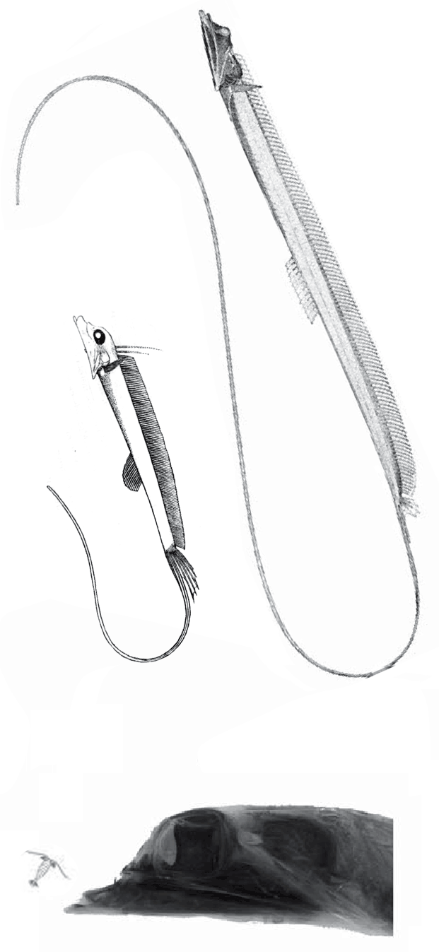
“The factors that prompted orbital reorganization in Thylacosmilus are unknown…” that may be because only one other ancestral taxon was listed in the text (house-cat sized Patagosmilus, Fig 2), but not illustrated. The authors could have answered this question by testing ancestral and related taxa. They did not do so. In the LRT this sabertooth marsupial clade goes back in time to rat-sized Early Cretaceous Vincelestes (Fig 2). Even deeper in this lineage are extant Dasycercus and Dasyuroides (Fig 6), also not mentioned in the text. The latter is known to, “kill with strong bites to the head” of its victims. Phylogenetic bracketing indicates that extant habit could help us understand the habits of the extinct related Thylacosmilus (Fig 1).

The authors > also < report,
“The forcing function behind these morphological tradeoffs was the extraordinary growth of its rootless canines, which affected skull shape in Thylacosmilus in numerous ways, including relative orbital displacement.”

So, “The factors that prompted orbital reorganization in Thylacosmilus” are known, according to the authors. This is easy to see (Fig 1). The question the authorsleave unanswered (due to taxon exclusion) is: how far back does this trait extend?

Figure 4. Patagosmilus and Vincelestes at full scale on a 72dpi monitor. These are derived members of the sabertooth marsupial clade.
Figure 2. Eocene Patagosmilus and Early Cretaceous Vincelestes at full scale on a 72dpi monitor. In the LRT these are members of the sabertooth marsupial clade that includes Thylacosmilus.

Gaillard, MacPhee and Forasiepi reported,
“We show that the orbits of Thylacosmilus were frontated and verticalized in a way that favored some degree of stereopsis and compensated for limited convergence in orbital orientation.

Figure 5. Skull of Barbourofelis, a traditional nimvravid placental cat, nests in the LRT with Thylacosmilus after trait analysis.
Figure 3. Skull of Barbourofelis, a traditional nimvravid placental cat, nests in the LRT with marsupial Thylacosmilus after trait analysis. Note the similar deep roots that rotate the orbits further laterally.

This can’t be the first time
this ‘limited convergence in orbital orientation’ has been noted. Or is it? Have these three authors finally unveiled the obvious? Or are they making a big deal out of something everyone is already aware of?

Figure x. Smilodon skull, skeleton, manus and pes with PILs added.
Figure 4. Smilodon skull, skeleton, manus and pes with PILs added. Compare to Stylinodon in figure y.

Barbourofelis
(Fig 3) is traditional nimravid (= placental cat), but in the LRT nests with the marsupial, Thylacosmilus (Fig 1), sharing a similar skull architecture not found in placental cats (including Nimravus Fig 5) or Smilodon, Fig 4). To force Barbourofelis to move in the LRT requires 28 additional steps to Smilodon, and 34 additional steps to Nimravus.

Figure 4. Osbornudon to scale with Nimravus.
Figure 5. The nimravid cats, Osbornudon to scale with Nimravus.

Phylogenetic analysis answers so many questions.
Build your own LRT so you can use this powerful tool to answer phylogenetic questions like those Gaillard, MacPhee and Forasiepi both posed and overlooked.

Figure 8. Extant Dasyuroides is a carnivorous marsupial in the lineage of Thylacosmilus.
Figure 6. Extant Dasyuroides is a carnivorous marsupial in the lineage of Thylacosmilus.

References
Gaillard C, MacPhee, RDE and Forasiepi AM 2023. Seeing through the eyes of the sabertooth Thylacosmilus atrox (Metatheria, Sparassodonta). Nature communications biology https://doi.org/10.1038/s42003-023-04624-5

wiki/Sparassodonta

Theropod lips? Don’t forget Sinornithosaurus.

“Animals like T-Rex, theropod dinosaurs, most likely had some sort of lips, like a soft tissue covering on their mouth to cover their teeth,” said one of the authors of the study, Thomas Cullen, an assistant professor of paleobiology at Auburn University.

As everyone knows, living theropods (birds) and living archosaurs (crocs) lack lips. On the other hand, lizards (including snakes) have lips, as do most tetrapods.

That sets the stage for this argument. Cullen et al 2023 decided to forsake phylogenetic bracketing and place their bets on convergence with lizards and their similar skull foramina.

Cullen et al also chose to illustrate their hypothesis
by using one Tyrannosaurus with relatively small teeth (Fig 1, FMNH PR 2081), rather than really test their hypothesis with several theropods with much larger lateral teeth (Figs 2, 4).

If Cullen et al were trying to set a rule, that rule should apply to one and all.
More toothy specimens (Figs 2, 4) were not illustrated in Cullen et al.

Figure 1. T- rex illustrations from Cullen et al 2023.
Figure 1. T- rex illustrations from Cullen et al 2023. Note the authors are indicating at least half of the upper tooth coverage comes from the lower lip. Their mismatch of the anterior dentary with the flesh reconstruction is forgivable, but indicates inaccuracy, leading to loss of confidence in the authors hypotheses, which should be flawlessly presented. Compare this skull with others in figure 2.

Cullen 2023 et al reported,
“Large theropod dinosaurs are often reconstructed with their marginal dentition exposed because of the enormous size of their teeth and their phylogenetic association to crocodylians. We tested this hypothesis using a multiproxy approach. Regressions of skull length and tooth size for a range of theropods and extant varanid lizards confirm that complete coverage of theropod dinosaur teeth with extraoral tissues (gingiva and labial scales) is both plausible and consistent with patterns observed in living ziphodont [= pointed, laterally compressed teeth, with serrated edges] amniotes.

Figure 2. Other specimens attributed to Tyrannosaurs animated to show tooth depth extends to the lower margin of the mandible or nearly so. Compare to the FMNH Tyrannosaurus in figure 1, which has relatively smaller teeth.

Cullen 2023 et al reported,
“Analyses of dental histology from crocodylians and theropod dinosaurs, including Tyrannosaurus rex, further indicate that the most likely condition was complete coverage of the marginal dentition with extraoral tissue when the mouth was closed.”

About those lower lip deep pockets purported to contain the upper teeth…
Cullen et al minimize their depth by permitting less jaw closure (Fig 3).

Figure 2. From Cullen et al 2023. Their cross-sections indicate an ability to close the jaws more when lips are missing AND contact between the tooth tips and lower lips.
Figure 3. From Cullen et al 2023. Their cross-sections indicate an ability to close the jaws more when lips are missing AND contact between the tooth tips and lower lips.

Key to their argument Cullen et al reported,
“the lower-density, linear pattern of foramina on the face and jaws of theropods,
such as tyrannosaurids, is as or more similar in structure to that of many extant squamates,
such as Varanus or Amblyrhynchus, than to the pattern observed in extant crocodylians
such as Alligator.”

For a rebuttal, see the Tracy Ford video cited below.

Figure 4. Skull of Sinornithosaurus updated. The large lateral maxillary teeth likely were exposed. If not, deeeeeep lip pockets would have been required. This image updates the one originally posted the day before by pushing the tooth roots back into their alveoli. The tooth tips no longer extend below the mandible ventral margin, but some extend to this ventral margin, still requiring deeep lip pockets.

Sinornithosaurus – a ‘worst-case scenario’
(Fig 4) pushes the Cullen et al hypothesis over the brink with its extra large lateral teeth that extend > below < the lower margin of the dentary whenever the jaws were closed… unless the jaws didn’t close due to shallow lip pockets, according to Cullen et al’s hypothesis.

Bottom line:
Pertinent taxa that might falsify the presented hypothesis were omitted from Cullen et al. Their study should have considered the ziphodont theropods with the largest lateral teeth if their hypothesis is indeed valid for all ziphodont theropods. The authors indicated no exceptions.

References
Carr et al 2017. A new tyrannosaur with evidence for anagenesis and crocodile-like facial sensory system. Scientific Reports 7:44942
Cullen TM et al 2023. Theropod dinosaur facial reconstruction and the importance of soft tissues in paleobiology, Science (2023). DOI: 10.1126/science.abo7877. www.science.org/doi/10.1126/science.abo7877

Tracy Ford on YouTube.
Scott Hartman writing on this subject reported the following:
“Bob Bakker was arguing for lizard-like lips on theropods in the pages of The Dinosaur Heresies back in the mid-1980s, an interpretation also championed by Greg Paul.”

“The authors of the D. horneri paper (Carr, et al.) looked closely at the texture of the bones of the skull and attributed a wide range of skin types to them, including keratinous horn-like material, and large and small scale patterns. Specifically relevant for us they interpreted the rugose patterns on the maxilla as being similar to the textured snouts of crocodilians. They, as well as Tracy Ford argue that the foramina (small holes for nerves and/or blood vessels) on the snout and jaws of tyrannosaurs may have made them with highly touch-sensitive, meaning that the nerve openings were to support tactile perception, not lips.”

“Non-bird theropods clearly have very different anatomy from from living crocodiles and alligators, in almost every way you can examine their oral and facial anatomy.”

Publicity
https://phys.org/news/2023-03-protruding-rex-teeth-lips.html

Additional publicity

The extant snakehead, Channa, has an Early Triassic relative, Watsonulus, a member of a traditionally extinct clade

The fish subset
of the Large Reptile Tree (LRT, 2223 taxa) is complete, but still subject to updates as they arise. This is one of the interrelationships that arose over the last several months of housekeeping.

Figure 1. The extant snakehead, Channa, compared to the related Watsonulus from the Early Triassic.
Figure 1. The snakehead, Channa, compared to the related Watsonulus from the Early Triassic, to scale.

Channa sp.
(Scopoli 1777; 25 cm to 1+m, Figs 1, 2) is the extant snakehead, a predatory freshwater fish nesting here with Watsonulus. This fish can breathe air and travel across land for short distances seeking new ponds, but this is rare as the pectoral fins are weak and poorly angled for this. The pelvic fins are absent. Teeth also are present on the parasphenoid. Nicknames include “Frankenfish” and “the fish from Hell”.

Figure 2. Left: Watsonulus skull. Right: Channa skull. Colors added here.

Watsonulus eugnathoides
(originally Watsonia Piveteau 1935; Olsen 1984; Early Triassic; 7cm skull length, 30cm long, Figs 1, 2) is a parasemionotid fish, traditionally thought to be an extinct clade. Note the large diamond opening between the nasals and frontals in dorsal view. The parietals are separated laterally by the postparietal. The prefrontal extends to produce a post-narial process. The teeth are extremely tiny. The pectoral fins are much larger than the pelvic fins.

References
Olsen PE 1984. The skull and pectoral girdle of the parasemionotid fish Watsonulus eugnathoides from the Early Triassic Sakamena Group of Madagascar, with comments on the relationships of the holostean fishes. Journal of Vertebrate Paleontology 4(3):481–499.
Piveteau J 1930. C R Acad. Sci. Paris, 191
Piveteau J 1935. Palkontologie de Madagascar. XXI. Les Poissons du Trias infkrieur. Contribution 2 l’ktude des actinoptkrygiens. Annales de Pal6ontologie 23:8 1-1 80.
Scopoli GA 1777. Introductio ad historiam naturalem, sistens genera lapidum, plantarum et animalium hactenus detecta, caracteribus essentialibus donata, in tribus divisa, subinde ad leges naturae. Pragae. Wolfgang Gerle. Pp i-x + 1-506.

wiki/Watsonulus
wiki/Channa
wiki/Snakehead
wiki/Parasemionotiformes

Beishanichthys updated

Phylogenetically
Early Triassic Beishanichthys (Figs. 1, 2) nests at the base of the ray fin fish clade (Actinopterygii) in the revised and growing large reptile tree (LRT, 2223 taxa). Beishanichthys is derived from more primitive palaeoniscid fish in the LRT.

Figure 1. Beishanichthys in situ.

Beishanichthys breviacaualis
(Xu and Gao 2011; Early Triassic) is a genus of scanilepiform fish nesting with Fukangichthys in the LRT at the base of the ray-fin fish. In some studies this clade is considered the most primitive basal bony fish.

Figure 2. Beishanichthys skull. Colors added here.
Figure 2. Beishanichthys skull. Colors added here. Note the tentative separation of the maxilla (green) from the lacrimal (tan), which originates far from the nose, but close to the gills.

Giles et al 2017 reported,
“We show that scanilepiforms [like Beishanichthys], a widely distributed Triassic (ca. 252-201 Mya) radiation, are stem polypterids.” And “Anatomical and molecular data now support placement of polypterids as the 48 living sister group of all other extant actinopterygians.”

These statements are not confirmed by the LRT, which nests Polypterus with lungfish, close to basal tetrapods, far from Beishanichthys.

Figure 3. Polypterus is a lungfish relative, not a Beishanichthys relative.

Housekeeping continues
in the fish subset of the LRT. Getting close now, but close still has problems. There is something to learn and discover every day because the problems encourage a deeper dive into the data (Figs 1, 2) to resolve those problems.

References
Giles S, Xu G-H, Near TJ; Friedman M 2017. Early members of ‘living fossil’ lineage imply later origin of modern ray-finned fishes. Nature. 549 (7671): 265–268.
Su T 1978. Memoirs Inst. Vert. Paleont. Paleoanthrop. Peking No. 13.
Xu G-H and Gao K-Q 2011. A new scanilepiform from the Lower Triassic of northern Gansu Province, China, and phylogenetic relationships of non-teleostean Actinopterygii PDF. Zoological Journal of the Linnean Society. 161 (3): 595–612.
Xu G-H, Gao K-Q and Finarell JA 2014. A revision of the Middle Triassic scanilepiform fish Fukangichthys longidorsalis from Xinjiang, China, with comments on the phylogeny of the Actinopteri. Journal of Vertebrate Paleontology 34(4):747–759.

wiki/Fukangichthys
wiki/Beishanichthys

Repairing a µCT scan of the deep sea slickhead, Narcetes

The fragile facial bones of ray-fin fish
are important when it comes to scoring them in the large reptile tree (LRT, 2223 taxa). So when they get jumbled, as in this cadaver (Figs 1, 2), it’s a good idea to use Photoshop to put the bones back to their original order and in vivo positions. Then score the traits.

To do this it with confidence takes a little experience to recognize previous errors and a good model, a Bauplan from a related taxon, to guide your changes.

Figure 1. µCT scan of the skull of the extant slickhead, Narcetes from Fujiwara et al 2021. Colors and facial bone shifts added here.

In this case the model for the 140cm long extant slickhead, Narcetes
(Fig 1) is a tiny 4cm long Middle Triassic basal bony ray-fin fish, Prohalecites (Fig 3). These two nest together in the LRT.

Figure 2. Narcetes from Fujiwara et al 2021. Compare to Prohalecites in figure 3.

Narcetes shonanmaruae
(Alcock 1890; Fujiwara et al. 2021; 140cm, typically 75cm) is the extant slickhead, a deep-sea relative of tiny Prohalecites. Anal fin entirely behind the dorsal fin, multiserial teeth on jaws, more scale rows than congeners, precaudal vertebrae less than 30, multiserial teeth on jaws, more scale rows than congeners, precaudal vertebrae less than 30, seven branchiostegal rays, two epurals, and head smaller than those of relatives. Most slickheads are benthopelagic or mesopelagic feeders of gelatinous zooplankton, but behavioural observations and dietary analyses indicate that the new species is piscivorous.

Figure 3. Tiny Middle Triassic Prohalecites nests with 1.4m long Narcetes in the LRT. Diagram from Tintori 1990.

Prohalecites porroi
Bellott 1857, Tintori 1990, MCSNIO P 348, Middle Triassic; 4cm) is a late surviving basal bony ray-fin fish (Actinopterygia, Osteichthyes). No trace of scales is preserved in any specimen (as in slickhead heads, Figs 1, 2). No neurocranial material is preserved. Tintori left Prohalecites as a Neopterygian incertae sedis, “because its characters do not perfectly fit in any of these cited groups.” Hemichordacentra are present. The preopercular is so slender it is rodlike.

Changes like this
(Fig 1) are only one reason why housekeeping on the ray-fin subset of the LRT is taking so long (moving from months to seasons).

This appears to be a novel hypothesis of interrelationships.
If not, please provide a citation so I can promote it here.

References
Alcock AW 1890. Natural history notes from H. M. Indian marine survey steamer ‘Investigator’, Commander R. F. Hoskyn, R. N., commanding. No. 18. On the bathybial fishes of the Arabian Sea, obtained during the season 1889-1890. [Unspecified Publisher] Vol. 6: 295-311.
Arratia G and Tintori A 1999. The caudal skeleton of the Triassic actinopterygian †Prohalecites and its phylogenetic position, p. 121–142. In: Mesozoic Fishes 2—Systematics and Fossil Record. G. Arratia and H.-P. Schultze (eds.). Verlag Dr. F. Pfeil, München.
Arratia G 2015. Complexities of early Teleostei and the evolution of particular morphological structures through time. Copeia 103(4):999–1025.
Bellotti C 1857. Descizione di alcune nuove specie di pesci fossili di Perledo e di altre localtta lombarde. 419–432. In Sopani A (ed) Studi geologici sulla Lomabardia. Editore Turati, Milano.
Fujiwara Y et al. 2021. Discovery of a colossal slickhead (Alepocephaliformes: Alepocephalidae): an active-swimming top predator in the deep waters of Suruga Bay,
Japan. Nature Scientific Reports 11:2490
Tintori A 1990. The actinopterygian fish Prohalecites from the Triassic of northern Italy. Palaeontology 33:155–174.

wiki/Prohalecites
wiki/Narcetes

A tiny, ancient, African river fish, Cromeria, is another odd sturgeon

Updated March 31, 2023
with new labels for certain bones in Cromeria (Fig 1), which allies it with Acipenser the sturgeon, and especially with its larvae (neotony at play, Fig 3). Recently a larger marine fish, Gonorynchus (Fig 3), also nested with sturgeons.

Cromeria nilotica
(Boulenger 1901; 4.5cm; Figs 1-3) is an extant tiny African naked shellear previously not associated with Gonorynchus and sturgeons. Note the subterminal mouth. The scaleless body lacks a lateral line. The caudal fin extends anteriorly both dorsally and ventrally.

Figure 2. From Gregory 1933, diagram of Cromeria. Colors added here. Note the odd caudal fin extending anteriorly to the dorsal and anal fins.
Figure 3. Two species of Cromeria, the African naked shellear shown several times life size.
Figure 2. Acipenser (sturgeon) larvae compared (not to scale) with an adult Gonorhynchus.

This appears to be a novel hypothesis of interrelationships.
lf not, please provide a citation so I can promote it here.

References
Boulenger GA 1901. Diagnoses of new fishes discovered by Mr. W. L. S. Loat in the Nile. Annals and Magazine of Natural History, Including Zoology, Botany and Geology, Being a Continuation of the ‘Magazine of Botany and Zoology’, and of Louden and Charlesworth’s ‘Magazine of Natural History’, Series 7 8: 444-446.

wiki/Shortnose_sturgeon_Acipenser
wiki/Gonorhynchus wiki/Gonorynchus_gonorynchus
wiki/Cromeria

With novel scoring Gonorynchus now nests with sturgeons in the LRT

Evolution, convergence, tradition and textbooks
made extant Gonorynchus (aka Gonorhynchus, Figs 1, 2) a difficult taxon to nest. Neotony (Fig 2) opened the door to possibilities. The large reptile tree (LRT, 2223 taxa) did the rest.

Evolution: No other fish have an underslung mouth, an operculum and a diphycercal tail.
Convergence: If this is not what the LRT indicates, then it’s more than superficially similar.
Tradition and textbooks: nest this taxon with dissimilar extant ‘relatives’.

Extant Gonorynchus gonorynchus
(originally Cyprinus gonorynchus Linneaus 1766; 60 cm, Figs 1, 2) goes by several common names including beaked salmon, ratfish, mousefish, sand eel, sand fish and shark whiting. The circumorbital bones are absent. The small, ventral jaw elements lack teeth. This nocturnal fish eats zooplankton and buried invertebrates, then buries itself during the day.

But what is it most closely related to? That’s the question.

Figure 1. Gonorhynchus nests by itself in most cladograms. New insights now nest it with sturgeons, based chiefly on skull traits.
Figure 1. Gonorhynchus nests by itself in most cladograms. New insights now nest it with sturgeons, based chiefly on skull traits illustrated by Gregory 1933. Colors added here. Note the label changes.

Gregory 1933 reported,
“It has an elongate cylindrical body and a sturgeon-like head with a pointed snout, a small inferiorly-placed mouth and a rostral barbel. The body and head are covered with small spiny scales.

Here several bones labeled in Gregory 1933 are relabeled
according to sturgeon homologies. Adult sturgeons likewise have no tooth-bearing mouth parts. The rostrum + barbels extend anterior to the ventral oral cavity. The mouth parts are protactile. A notochord pierces the well-ossified abdominal vertebrae. The torso is squarish in cross-section. The pectoral fins are essentially immobile, all as in sturgeons.

Figure 2. Acipenser (sturgeon) larvae compared (not to scale) with an adult Gonorhynchus.
Figure 2. Acipenser (sturgeon) larvae compared (not to scale) with an adult Gonorhynchus. Adult Acipenser lose the premaxilla, dentary and their ‘teeth’. Those calcium apatite ‘teeth’ are inherited from lamprey, hagfish and conodont ancestors. Enamel teeth appear later among placoderms. Not sure yet how those oral elements move.

As in sturgeon larvae,
(Fig 2) a premaxilla and preopercular are present. A postorbital is absent. The prefrontal is separated from the postfrontal.

Figure 2. Acipenser brevirostrum, 1m typical length. Records up to 1.47m.
Figure 3. Acipenser brevirostrum, 1m typical length. Records up to 1.47m.

Distinct from an adult sturgeon
(genus: Acipenser, Fig 3), a jugal is absent in Gonorynchus (Fig 1). The tail is diphycercal. Armor plates are absent. The prefrontal is separate from the postfrontal.

Sturgeon ancestors
in the LRT go back to the Early Cambrian. The armor plates are vestiges of ancestral full body armor, as seen in Osteostraci like Hemicyclaspis. Extant sturgeons are a diverse lot with some taxa having a longer preorbital than postorbital skull, as in Gonorynchus.

Are those tiny toothless oral elements in Gonorynchus original equipment handed down with few changes from sturgeon ancestors? Or are those vestigial jaws that once had enamel teeth in some other ancestors? That’s the other question.

This appears to be a novel hypothesis of interrelationships.
If not, please provide a citation so I can promote it here. This hypothesis now requires confirmation, refutation or modification from other workers. Earlier (May 2022) the LRT nested Gonorynchus with bonefish, but noted the many surgeon traits. Corrections are part of the process of science.

PS
I recently became aware of the more intrusive, larger and more numerous advertising that has invaded this blogpost. Apologies. Apologies. Apologies.

References
Gregory WK 1933. Fish skulls. A study of the evolution of natural mechanisms. American Philosophical Society 23(2). Republished by Eric Lundberg, Laurel, Florida 1959.
Linneaus C von 1766. Systema naturæ per regna tria naturæ, secundum classes, ordines, genera, species, cum characteribus, differentiis, synonymis, locis. Tomus I. Editio duodecima, reformata. pp. 1–532. Holmiæ. (Salvius)

wiki/Gonorynchus
wiki/Gonorynchus_gonorynchus

Cheating anatomy to promote the ‘deer-like ancestry of whales’ hypothesis

Philip Perry, writing for BigThink.com
referenced a five-year-old paper by Vermeij and Motani 2018 regarding land-to-sea transitions in vertebrates. Perry’s online article was promoted with a photo of a model of Indohyus and the caption “Note it’s deer-like feet.” Unfortunately, the fossil of Indohyus does not have deer-like feet (Fig 1). As you can see, the feet of Indohyus are actually quite broad with spreading toes, like those of Pakicetus (Fig 2). Cheating anatomy is not good for science.

Figure 1. Model of Indohyus from the BigThink.com article compared to fossil material of Indohyus.

Promoting the deer-like hypothesis, the authors of Wikipedia wrote:
“Indohyus is an extinct genus of digitigrade even-toed ungulates known from Eocene fossils in Asia. This small chevrotain-like animal found in the Himalayas is one of the earliest known non-cetacean ancestors of whales.”

That hypothesis of interrelationships was shown to be false in 2016 due to taxon exclusion. It also does not make sense – but is widely embraced at the university level. According to the large reptile tree (LRT, 2223 taxa) Indohyus nests with an extant taxon, Tenrec (Fig 2), not with ungulates.

Figure 3. Skeleton of Tenrec alongside restored skeleton model of Pakicetus.
Figure 2. Skeleton of Tenrec alongside restored skeleton model of Pakicetus. Note: neither has deer-like feet. Neither are herbivores.

This all goes back to Gingerich et al 2001 and Thewissen et al 2007,
who wrote about “aquatic artiodactyls”. See figure 3.

Figure 4. Image from BigThink.com showing Elomeryx instead of Indohyus basal to whales in general.
Figure 3. Image from BigThink.com substituting Elomeryx instead of Indohyus basal to whales in general. This is a false narrative promoted by university professors and the press.

This whale origin problem could have been corrected several years ago,
but workers refereeing a manuscript submission rejected it. The manuscript points to ‘taxon exclusion’ as the only reason prior workers missed the mark. That might have been embarrassing to the academics like Gingerich and Thewiessen who mistakenly promoted the ‘early artiodactyl‘ and ‘aquatic artiodactyl’ hypothesis.

This instance of rejection was not an isolated incident, but is a common practice in paleontology. That’s why it took over a century for paleontologists to embrace the ‘birds are dinosaurs’ hypothesis and why other workers continue to embrace the ‘bat-wing bird’ hypothesis, among a slew of other bogus hypotheses presented to tuition-paying students.

You can read that rejected manuscript by Googling “Triple origin of whales”, at ResearchGate.net.

Special thanks to paleontologist, Don Prothero,
who linked the BigThink.com article to his Facebook page.

References
Gingerich, PD, Haq M, Zalmout IS, Khan IH, and Malkani MS 2001. Origin of whales from early artiodactyls: hands and feet of Eocene Protocetidae from Pakistan. Science, 293:2239-2242.
Perry P 2023. Ancient deer-like creatures returned to the ocean to become whales. But why? BigThink.com
Thewissen JGM, Cooper LN, Clementz MT, Bajpai S and Tiwari BN 2007. Whales originated from aquatic artiodactyls in the Eocene epoch of India. Nature 450:1190-1195.
Vermeij GJ and Motani R 2018. Land to sea transitions in vertebrates: the dynamics of colonization. Paleobiology 44(2):237–250. online here

wiki/Indohyus

Tiny, toothless Stylephorus now nests with another blenny: toothy Gigantura

Untested traits include
those cylindrical eyeballs and that elongate lower caudal fin.

Figure 1. Gigantura compared to Stylephorus to a reduced scale.

Gigantura indica
(Brauer A 1901, Konstantinidis and Johnson 2016; 20 cm standard length, not counting the caudal fin, Figs 1–3) is the extant telescope fish. Comparisons with sister taxa indicate the indicated ‘palatine’ with teeth is actually the premaxilla in figure 2 from Gregory 1933. The toothless maxilla (green) is located at the posterior mandible. The postorbital (=circumorbital ring) is reduced to two vestiges. This small fish can swallow prey larger than itself.

Figure 2. Skull of Gigantura from Gregory 1933. Colors added here.
Figure 2. Skull of Gigantura from Gregory 1933. Colors and new labels added here.
Figure 3. Gigantura magnified.
Figure 3. Gigantura magnified to show facial details including the cylindrical eyeballs.

Stylephorus chordatus
(Shaw, 1791, Regan 1924, Figs 4, 5) is the extant tube-eye or thread-tail. It was considered an oarfish relative (Fig 6), but here nests with Gigantura (Figs 1–3). Note the large eyes and flexible neck. Convergent with seahorses and oarfish, the tube-eye also feeds on tiny plankton sucked in as its tubular mouth enlarges the oral cavity by 40x (Figs 4, 5).

Figure 4. Stylephorus skull animated. Colors added here.
Figure 4. Stylephorus skull animated. Colors added here. Compare to Gigantura in figure 1. The maxilla (mx) labeled here is actually the anterior portion of the postorbital. And now you know why ray-fin fish can be difficult to score.
Figure 1. Stylephorus ontogeny and feeding
Figure 5. Stylephorus ontogeny. Feeding animated. Much enlarged. Note the eye-cylinders.

Both of these odd deep-sea fish
find a last common ancestor in the large reptile tree (LRT, 2223 taxa) close to Acanthemblemaria, the tube blenny (Fig 7). So all three are blennies.

Figure 7. Traditional fish cladogram nesting Stylephorus with opahs and oarfish.

Acanthemblemaria aceroi
(genus: Metzelaar 1919; species: Hastings, Eytan and Summers 2020) is a newly described tube blenny described with a µCT scan skeleton. Note the vestige maxilla (green) detached from the lacrimal (tan). Here it nests with Neoclinus, the sarcastic fringhead blenny.

Figure 7. Acanthemblemaria, the tube blenny is presently the last common ancestor of Gigantura and Stylephorus in the LRT. Note the emphasis on the eye sockets and the diminution of the maxilla (green) with a large vestige posteriorly, as in Gigantura (Fig 1).

Housekeeping the ray-fin fish clade continues unabated,
testing taxa together that rarely get tested together in trait analysis.

References
Brauer A 1901. Über einige von der Valdivia-Expedition gesammelte Tiefseefische und ihre Augen. Sitzungsberichte der Gesellschaft zur Beförderung der Gesamten Naturwissenschaften zu Marburg 8: 115–130.
Konstantinidis P and Johnson GD 2016. Osteology of the telescope fishes of the genus Gigantura (Brauer, 1901), Teleostei: Aulopiformes. Zoological Journal of the Linnean Society 179(2):338–353.
Regan CT 1924. The morphology of the rare oceanic fish, Stylophorus chordatus, Shaw; based on specimens collected in the Atlantic by the “Dana” expeditions, 1920–1922. Proceedings of the Royal Society B 96(674): PDF
Shaw G 1791. Description of the Stylephorus chordatus, a new fish. Transactions of the Linnean Society of London, 2d Ser: Zoology 1:90–92.

wiki/Stylephorus
wiki/Sarcastic Fringehead
wiki/Acanthemblemaria
wiki/Gigantura

Opah relatives in the LRT

Recent housekeeping
on the ray-fin fish portion of the large reptile tree (LRT, 2223 taxa) is finally coming to a conclusion (let’s hope!). Today’s graphic (Fig 1) presents the large, disc-shaped opah (genus: Lampris) and a menagerie of equally odd close relatives. These include the surface sprinter, Exocoetus, the deep-sea flashlight fish, Anomalops, and the elephant-nosed Gnathonemus (Fig 1). The sole fossil taxon, Massamorichthys, is from the Paleocene.

Figure 1. Lampris, the opah, shown in two views, plus its many smaller relatives to scale. Presently the last common ancestor of Lampris and Seriola rivoliana is the tiny stickleback, Gasterosteus, the outgroup taxon, which in turn is basal to pipefish, oarfish and seahorses.