
Figure 1. Tiarajudens newly reconstructed. Here it appears that the tusk is not a canine, but the last maxillary tooth, posterior to the apex of the maxilla, distinct from all other therapsids and distinct from dicynodonts, with whom Tiarajudens has been nested in prior studies.
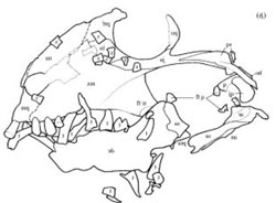
Figure 2. Anomolocephalus. Are those flat teeth, or broken. If flat they are autapomorphic, unlike sister taxa. If elongated, they are synapomorphies, like sister taxa.

Figure 2. Anomocephalus skull with teeth replaced, no longer loose. It is apparent that the teeth ground against one another.
Sorry for the lack of posts in the last two days.
This is what I’m working on. The therapsid tree is going to change a little. More tomorrow.
Change made Nov. 16, 2013
The skull of Anomocephalus is more accurately reconstructed below (Fig. 3) based on Modesto and Rubidge 2000.
References
Modesto S and Rubidge B 2000. A basal anomodont therapsid from the lower Beaufort Group, Upper Permian of South Africa. Journal of Vertebrate Paleontology 20(3):515-521.

Why is the break in the maxilla/premax area of Tiarajudens so much larger in your reconstruction than in the actual specimen? What evidence is there to suggest displacement there?
Nice, high-res photo: http://upload.wikimedia.org/wikipedia/commons/a/ae/Tiarajudens_eccentricus_skull.jpeg
In your reconstruction the tusk does not appear to be the “last maxillary tooth” as you stated – there certainly looks like teeth on either side of the tusk in your reconstruction.
I know I’m probably getting ahead of myself since you said more details are coming…