
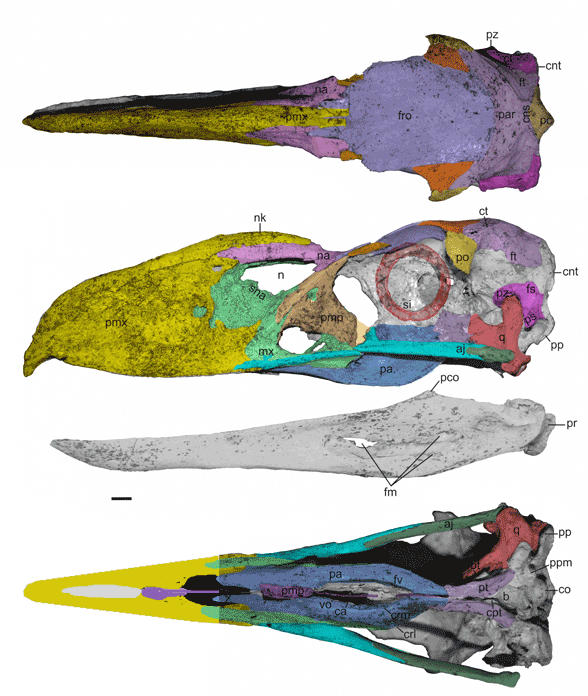
Figure 3. Llallawavis skull in 3 views with DGS identifying bones, many of which are fused to one another. Image from DeGrange et al. with DGS added. Note DeGrange et al. labeled bones but did not delineate sutures. That needs to be done. Here the palatine is shown to be restricted to a small area below the maxilla and the rest is the pterygoid. Note the expansion of the maxilla in the reinforcement of this killing beak.
Figure 3. Llallawavis skull in 3 views with DGS identifying bones, many of which are fused to one another. Image from DeGrange et al. with DGS added. Note DeGrange et al. labeled bones but did not delineate sutures. That needs to be done. Here the palatine is shown to be restricted to a small area below the maxilla and the rest is the pterygoid. Note the expansion of the maxilla in the reinforcement of this killing beak.
